
Medical Global Academy

August 20, 2026

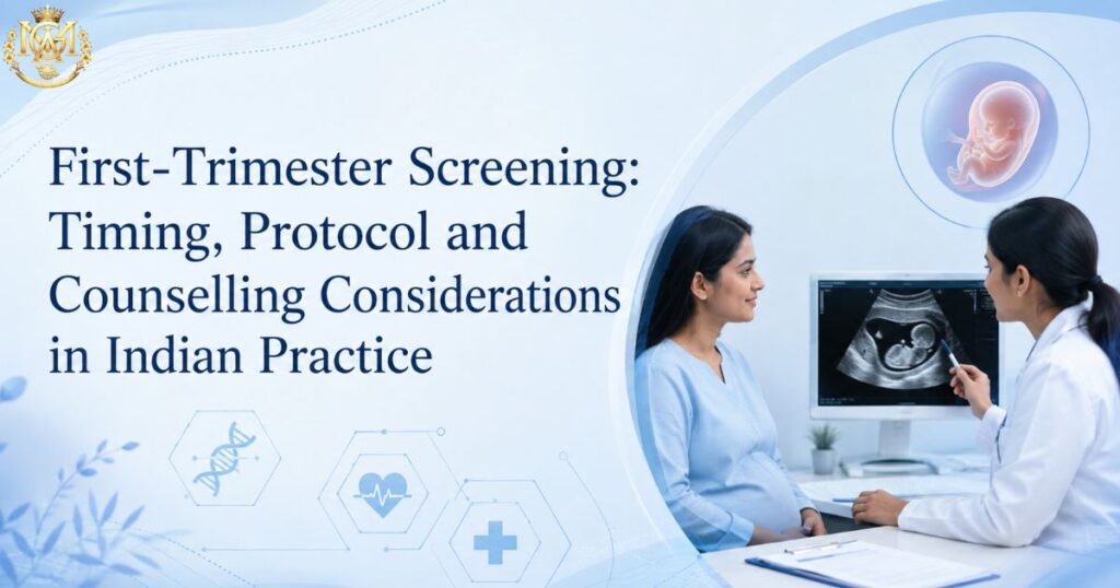
Doctor explaining first-trimester screening protocols and NT scan results to a patient
Learning blogs

First-Trimester Screening: Timing, Protocol and Counselling Considerations in Indian Practice

Quick Answer First-trimester screening for chromosomal abnormalities is optimally performed between 11+0 and 13+6 weeks of gestation. The combined screening test — NT measurement plus maternal serum free β-hCG and PAPP-A — achieves detection rates of approximately 85–90% for Down syndrome (Trisomy 21) at a 5% false-positive rate when performed within this window. NIPT (Non-Invasive Prenatal Testing) can be offered from 10 weeks onwards and carries a higher sensitivity, but does not replace diagnostic confirmation in screen-positive cases. This article covers the timing rationale, measurement standards, counselling framework, and the specific considerations that apply in Indian practice. First-trimester screening is one of the most clinically consequential steps in antenatal care — and one of the areas where clinical practice among OBGYN doctors varies most. Timing errors, suboptimal NT measurement technique, incomplete counselling, and inconsistent integration of combined screening markers are all common in routine practice, and all affect the clinical utility of the screening result. This article is a protocol-level reference. It covers the timing window and why it matters, the components of combined screening, NT measurement standards, the role of NIPT, second-line biochemical markers, and the counselling framework a practising clinician needs when delivering a screen-positive result. It also addresses the specific legal and procedural context that applies to prenatal diagnosis in India. Doctors who want structured training in prenatal screening interpretation and fetal ultrasound can explore MGA’s Fellowship in Fetal Medicine, which covers this content in depth across dedicated modules. 11–14 wk Optimal combined screening window 85–90% T21 detection rate — combined screening >99% NIPT sensitivity for T21 What this article covers Why the 11–14 week window matters — the biological rationale NT measurement — standards and common errors Combined first-trimester screening — components and interpretation NIPT — role, timing, and limitations in Indian practice Second-trimester integration — when to offer the quadruple test Screen-positive results — the counselling framework PCPNDT context — what practitioners need to know Frequently asked questions Why the 11–14 week window matters — the biological rationale The 11+0 to 13+6 week window is not arbitrary. Nuchal translucency — the fluid-filled space at the back of the fetal neck, measured on ultrasound — reaches its maximum diagnostic utility within this window. NT is physiologically present in all fetuses to a small degree, and naturally increases after 14 weeks in unaffected pregnancies as lymphatic drainage establishes. Outside this window, NT measurements are either technically not yet feasible (before 11 weeks, the fetus is too small for reliable measurement) or diagnostically unreliable (after 14 weeks, NT has already begun to resolve in normal fetuses, narrowing the gap between affected and unaffected). The CRL (crown-rump length) range within this gestational window is 45–84 mm. The CRL measurement determines whether the fetus is within the NT measurement window, not gestational age alone — this distinction matters when dates are uncertain. Practical point: When a patient presents at or near 14 weeks, do not delay referral to a better-equipped centre if your local equipment or training is not adequate for NT measurement. A NT measurement at 13+5 is still within protocol; a measurement at 14+1 is not. Missing the window means the combined test cannot be performed and the patient defaults to second-trimester screening alone, which has lower sensitivity. NT measurement — standards and common errors NT measurement is the most skill-dependent component of first-trimester screening. Poorly performed NT measurements produce unreliable risk calculations regardless of how good the serum marker results are. Measurement standards (FMF protocol) The Fetal Medicine Foundation (FMF) protocol is the widely used international standard. Key requirements: Fetal position: Sagittal section of the fetus in a neutral position — neither hyperflexed nor hyperextended. Both hyperflexion and hyperextension artificially alter the NT measurement. Image magnification: The fetal head and upper thorax should occupy the full screen. The fetus should fill at least 75% of the screen. Calipers: Placed on the inner borders of the nuchal space — not on the skin lines themselves. The horizontal part of the caliper should be barely visible against the echogenic line. Skin line vs amnion: The amniotic membrane must not be confused with the skin line. Wait for the fetus to move away from the amnion if they appear fused. Multiple measurements: Take at least three measurements and use the largest for risk calculation. Common measurement errors Error Effect on Measurement Correction Neck hyperflexion Falsely increases NT by up to 0.6 mm Reposition, wait for neutral posture Measuring on amnion, not skin Falsely increases NT Wait for fetal movement to separate amnion Calipers on outer borders Overestimates NT Use inner-to-inner caliper placement Insufficient magnification Imprecise caliper placement Head and thorax to fill >75% of screen Single measurement accepted Misses higher reproducible value Minimum 3 measurements; use the largest Combined first-trimester screening — components and interpretation Combined screening integrates three inputs into a single risk calculation for chromosomal abnormalities, primarily Trisomy 21 (Down syndrome), Trisomy 18 (Edwards syndrome), and Trisomy 13 (Patau syndrome). Three components 1. Maternal age-related background risk: The prior probability of chromosomal abnormality based on the mother’s age at the expected date of delivery — the starting point before any test modifies it. 2. Nuchal translucency (NT) measurement: Expressed as a multiple of the median (MoM) for that gestational age and CRL. An NT at or above the 95th centile (approximately ≥2.5 mm at 11 weeks, ≥3.5 mm at 13 weeks) raises the posterior risk substantially, though the precise risk change depends on the full calculation. 3. Maternal serum markers — free β-hCG and PAPP-A: Free β-hCG is elevated in Down syndrome pregnancies at 11–13 weeks (approximately 2.0 MoM). PAPP-A is reduced in Down syndrome pregnancies at 11–13 weeks (approximately 0.4–0.5 MoM). These two markers change in opposite directions in Trisomy 21, which is what gives the combined test its discriminatory power. Risk threshold and result interpretation The standard combined screening risk threshold for classifying a result as screen-positive is 1 in 300 (or 1:300) for Trisomy 21. Results above this threshold (e.g., 1:50, 1:120) are screen-positive;
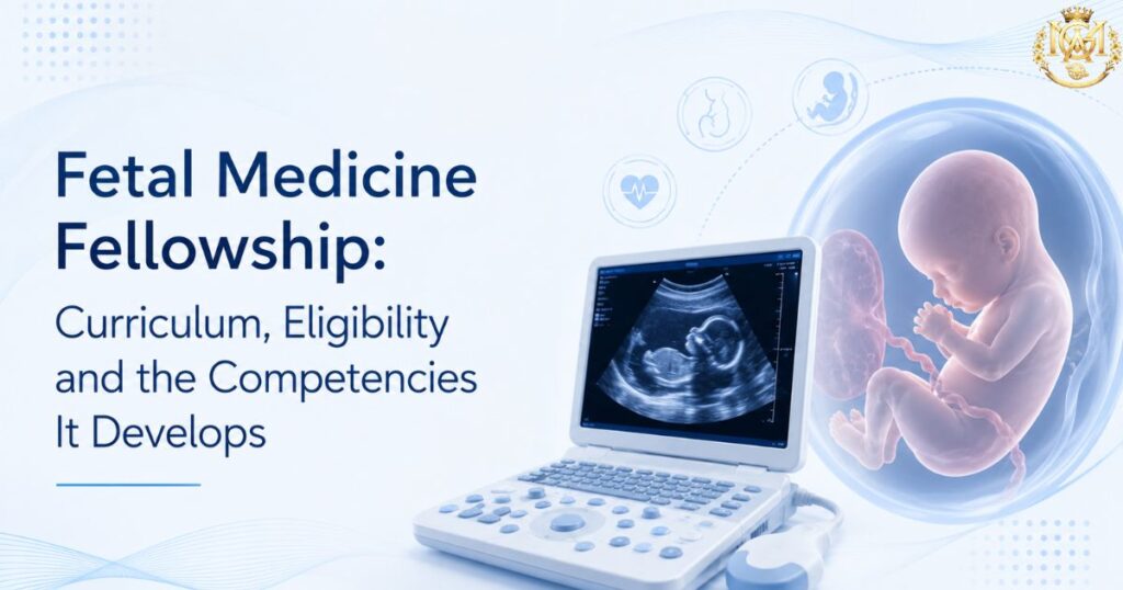
Fetal Medicine Fellowship curriculum and eligibility guide next to an ultrasound machine
Learning blogs

Fetal Medicine Fellowship: Curriculum, Eligibility and the Competencies It Develops

Quick Answer MGA’s Fellowship in Fetal Medicine is a 12-month, CPD-accredited online program covering 17 curriculum modules across fetal ultrasound, prenatal screening, fetal echocardiography, invasive procedures, high-risk pregnancy management, fetal anomaly detection, and prenatal genomics. It is designed for doctors with an obstetrics and gynaecology background — DGO, MD/MS OBG, and DNB OBGYN — who want structured subspecialty training in fetal medicine and prenatal diagnosis without pausing clinical practice. This article covers what the program specifically teaches, who is eligible, what procedural competencies it develops, and how it differs from general OBG training. If you are already practising obstetrics and considering a structured subspecialty commitment in fetal medicine, you have probably already read the persuasion-framed articles — the ones that explain what fetal medicine is and why it matters. This article serves a different purpose. It covers the specification: what MGA’s Fellowship in Fetal Medicine actually teaches module by module, who is eligible to apply, what procedural competencies it develops, and how to evaluate whether this program fits your current clinical context and career direction. For the case for why fetal medicine subspecialisation makes clinical sense for practising OBGYN doctors, MGA’s existing article on why to consider a fellowship in fetal medicine covers that well. This article picks up where that one leaves off. 17 modules Full curriculum breadth 12 months Duration — fully online CPD Accredited — international recognition What this article covers Program format and delivery — how it works for working doctors Eligibility — who the program is designed for 17-module curriculum — what is taught and in what sequence Core procedural competencies developed What this fellowship does not cover — honest scope setting How the fellowship positions you in subspecialty practice Frequently asked questions Program format and delivery — how it works for working doctors The fellowship runs for 12 months and is delivered entirely online — live sessions scheduled after OPD hours, with 100% recorded lectures available on the LMS portal for any session you cannot attend live. This format is intentional: it is built for doctors who are actively practising and cannot pause clinical work for a full-time program. The program includes weekly case clinic discussions, dedicated Q&A sessions, free study materials and an e-library with prenatal imaging guides and case references, CPD-accredited certification on completion, and one year of structured expert mentorship after the program ends. The mentorship period is not a nominal credential — it is designed to support your transition into applied fetal medicine practice once the academic year is complete. Accreditation: The fellowship is accredited by the CPD & Training Council with international continuing medical education recognition. It does not carry a university degree credential — it is a CPD-accredited professional fellowship. For doctors who specifically need a university-awarded qualification in obstetric imaging, MGA’s PG Diploma in Ultrasonography or Certificate in Advanced Ultrasound in Obstetrics and Gynecology are separate, differently-credentialled options. Eligibility — who the program is designed for The fellowship is open to eligible medical graduates. Based on the program’s clinical orientation — prenatal diagnosis, fetal ultrasound interpretation, invasive procedures, and high-risk pregnancy management — it is most directly relevant to doctors with an obstetrics and gynaecology background. Qualification Clinical Relevance to This Fellowship DGO Direct application — DGO doctors managing antenatal OPDs benefit most from structured fetal ultrasound, anomaly screening, and prenatal counselling modules MD / MS Obstetrics & Gynaecology Strong fit — builds subspecialty depth in fetal imaging, invasive procedures, and high-risk pregnancy beyond the MD/MS curriculum DNB OBGYN Strong fit — same clinical scope as MD/MS OBG; fellowship adds focused fetal medicine competency MBBS with OBG caseload Applicable — particularly for MBBS doctors working in antenatal care or managing obstetric patients in primary or secondary care settings General Physicians managing antenatal patients Relevant for the prenatal screening, high-risk pregnancy, and referral threshold modules — less directly applicable to the procedural content Check the fellowship course page for current eligibility requirements before applying — criteria may be updated periodically. 17-module curriculum — what is taught and in what sequence The 17 modules are sequenced to build from foundational knowledge through clinical application and into advanced and emerging practice areas. The outline below summarises each module’s scope. Module 1: Introduction to Fetal Medicine and Maternal-Fetal Physiology The foundational module establishes the clinical role of fetal medicine in contemporary obstetrics, covering maternal-fetal physiology, placental development and function, fetal growth, embryology, and an introduction to prenatal screening and genetic counselling frameworks. Module 2: Ultrasound and Imaging Techniques in Fetal Medicine Practical fetal ultrasound — guidelines for obstetric and gynaecological imaging, fetal biometry and growth assessment, Doppler ultrasound for fetal wellbeing, 3D/4D imaging of fetal abnormalities, amniotic fluid index assessment, placental and umbilical cord evaluation, and the application of ultrasound in chromosomal abnormality screening. Module 3: Prenatal Screening and Diagnosis First- and second-trimester aneuploidy screening protocols, NIPT clinical application and interpretation, combined screening markers, ultrasound indicators for Down syndrome and Trisomy 18, invasive diagnostic procedures (amniocentesis, CVS, fetal blood sampling), and genetic counselling communication with patients. Module 4: High-Risk Pregnancy and Maternal-Fetal Medicine Management of preeclampsia and gestational diabetes, hypertensive disorders of pregnancy including HELLP syndrome, fetal growth restriction and IUGR, multiple pregnancy diagnosis and management, premature labour prevention, antenatal corticosteroid therapy, and evidence-based monitoring of high-risk pregnancies. Module 5: Fetal Anomalies and Intervention Common fetal malformations — neural tube defects, congenital heart disease, gastrointestinal and renal defects — alongside fetal intervention indications, techniques, and outcomes, intrauterine fetal transfusion, post-fetal surgery neonatal care, and the ethical dimensions of fetal anomaly management. Module 6: Maternal-Fetal Medicine Procedures and Interventions Cordocentesis and fetal blood sampling, intrauterine treatments including laser therapy, fetal blood transfusion, and shunts, amniocentesis and CVS technique, indication, and risk counselling, and fetal wellbeing monitoring through biophysical profiles. Module 7: Ethical and Legal Aspects of Fetal Medicine Ethical dilemmas in fetal medicine practice, fetal rights versus maternal rights, decision-making frameworks for complex fetal conditions, informed consent for genetic counselling and invasive procedures, and the legal context of prenatal diagnosis in

Scroll to Top