
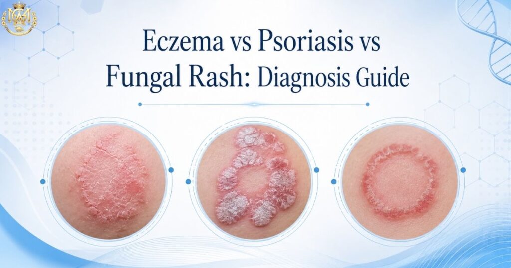
Eczema vs Psoriasis vs Fungal Rash: A Differential Diagnosis Guide for General Physicians
🩺 DIFFERENTIAL DIAGNOSIS MGA Clinical Education Series · 9 min read Eczema vs psoriasis vs fungal rash — three of the most commonly confused presentations in general practice — can look nearly identical at first glance. A patient walks into your clinic with an itchy, red, scaly rash on the elbows and knees… (rest of paragraph unchanged)A patient walks into your clinic with an itchy, red, scaly rash on the elbows and knees. Another presents with widespread, poorly-defined pale patches on the trunk after using an OTC cream. A third has a raised, silvery-scaled plaque on the extensor surface of the forearm that has been there for months. Three patients, three different conditions — but from a cursory clinical glance, they can look confusingly similar. Misdiagnosis between eczema (atopic dermatitis), psoriasis, and tinea (fungal rash) is remarkably common in general practice globally — and even more common in India, where steroid-modified tinea has become an epidemic masquerading as other conditions. This eczema vs psoriasis vs fungal rash guide walks through the key clinical differentiators that help a general physician reach the correct diagnosis quickly in the OPD, with a side-by-side comparison table and a simple decision framework that works in real-world practice. ⚡ Quick Differential Summary Eczema: Itch comes first → redness follows from scratching. Flexural surfaces (elbows, knees, neck). Lichenified skin, poor demarcation. Psoriasis: Well-demarcated plaques with silvery scale. Extensor surfaces (elbows, knees, shins, scalp). Systemic features possible (nail pitting, arthritis). Fungal (Tinea): Ring-shaped or atypical borders. Active border + central clearing (or pale centre if steroid-modified). KOH positive. Localized itch at border. Red flag: History of OTC cream use → suspect steroid-modified tinea mimicking eczema or psoriasis. 🔀 Why Eczema vs Psoriasis vs Fungal Rash Get Confused So Often All three conditions produce erythema (redness), scaling, and pruritus (itching). Each can present as a localized patch or become widespread, and each follows a chronic course. To an untrained eye — and occasionally even to a trained one — they appear nearly identical. The reason for confusion is structural: eczema and psoriasis are conditions of altered cutaneous immunity and barrier function; tinea is an infection. These arise from entirely different mechanisms — but the endpoint (inflammation, scaling, itch) is visually similar. Add to this the reality that in India, a fungal infection that has been treated with steroids (steroid-modified tinea) can look exactly like eczema or psoriasis, and misdiagnosis becomes not just common but expected. A machine-learning study published in Frontiers in Medicine (Ding et al., 2025, doi: 10.3389/fmed.2025.1667794) found that even with routine clinical data, eczema and psoriasis could not be distinguished with complete accuracy — illustrating that this differential is nuanced even with systematic approaches. The addition of a third condition (tinea) adds complexity but also opportunity: because tinea is fundamentally an infection, it responds to antifungals and has specific diagnostic tests (KOH), whereas eczema and psoriasis do not. 🧴 Eczema (Atopic Dermatitis): Key Clinical Features Classic phrase: “The itch that rashes” — not “the rash that itches.” This is the most clinically useful way to remember eczema’s hallmark. The itching comes first, often severe; scratching produces the visible rash and inflammation. 🔍 Eczema: Clinical Checklist Distribution: Flexural surfaces — antecubital fossa (crook of elbow), popliteal fossa (back of knee), neck, face, hands. In adults, often limited to hands or face. Children present with more widespread involvement. Appearance: Poorly demarcated (ill-defined borders). Lichenification (thickened, leather-like skin from chronic scratching). Erosions from scratching. Acute phase: vesicles and weeping. Chronic phase: dry, cracked, thickened skin. Itch character: Intense, often worse at night. Described as “maddening.” Scratch-itch cycle is central to perpetuation — the more you scratch, the more inflamed it becomes, the more it itches. Associated features: History of atopy (allergic asthma, allergic rhinitis, food allergy). Dry skin (xerosis) as baseline. May respond temporarily to steroids but relapses when stopped — if steroids alone are used without addressing barrier dysfunction. Response pattern: Responds well to emollients (moisturisers) + topical corticosteroids for acute flares + long-term barrier repair. Once correctly identified, it is predictable and manageable. 🏛️ Psoriasis: Key Clinical Features Classic phrase: “The rash that (sometimes) itches” — the plaque is present from the outset; itch is secondary and variable. An autoimmune condition in which the immune system attacks skin cells, causing rapid epidermal turnover. 🔍 Psoriasis: Clinical Checklist Distribution: Extensor surfaces — elbows, knees, shins, forearms, scalp, lower back, nails, ears. Psoriasis “loves bones” (bony prominences). Face and flexural areas are usually spared, which helps differentiate from eczema. Appearance: Well-demarcated plaques with a silvery or mica-like scale. The scale is often thick and adherent — when scraped off, pinpoint bleeding occurs (Auspitz sign). Sharp borders are characteristic. Lesions are thicker and more planar than eczema. Itch character: Variable — some patients report no itch at all; others report burning or soreness rather than intense itch. The plaque is visible and persistent; it does not improve simply from avoiding scratching. Associated features: Family history (positive in ~30% of patients). Nail involvement is common — pitting (tiny holes), onycholysis (separation from nail bed), subungual hyperkeratosis. Some patients have psoriatic arthritis (joint inflammation) — a key systemic clue not present in eczema or tinea. Response pattern: Chronic, cyclical (periods of remission and flare). Responds to topical steroids and vitamin-D analogues. Systemic triggers include stress, infections, certain medications (beta-blockers, lithium). Unlike tinea, psoriasis is never eradicated — it is managed long-term. 🍄 Fungal Rash (Tinea): Key Clinical Features Classic phrase: “Ring-shaped with a moving border.” The active border (periphery) is the site of active fungal invasion; the centre may clear (classic ringworm) or remain pale and poorly-defined if the patient has applied a steroid (steroid-modified tinea). 🔍 Fungal Rash (Tinea Corporis/Cruris/Faciei): Clinical Checklist Distribution: Any site — trunk, flexural and extensor surfaces equally affected. Tinea corporis affects the trunk and limbs, while tinea cruris involves the inguinal fold, pubic area, and inner thigh — the moist areas fungi favour. Tinea faciei, involving the face, is less common but increasing. Appearance: Classic: