

Medical Global Academy

September 4, 2026

Cardiologist reviewing an echocardiogram representing pathophysiology of heart failure and cardiomyopathies
Learning blogs

Heart Failure and Cardiomyopathies: Understanding the Heart’s Struggle to Keep Up

Cardiovascular Health A clear guide to pathophysiology, neurohormonal remodeling, genetics, and modern management — from first-line medication to transplant. In This Guide Pathophysiology of Heart Failure Management of Heart Failure Cardiomyopathies: The Four Major Types A Condition That Rewards Early, Coordinated Care Heart failure isn’t a single disease — it’s the endpoint of many different paths the heart’s muscle, valves, or rhythm can take when something goes wrong. Understanding heart failure and cardiomyopathies together matters, because cardiomyopathies are among the most important underlying causes of heart failure itself. At its core, heart failure means the heart can no longer pump enough blood to meet the body’s needs, or can only do so at abnormally high filling pressures. Cardiomyopathies — diseases that begin in the heart muscle itself — explain why treatment for heart failure and cardiomyopathies today looks so different from a decade ago. For doctors looking to build deeper expertise in this area, MGA’s Fellowship and PG Diploma programmes offer structured, CPD-accredited training across cardiovascular and related specialties. Pathophysiology of Heart Failure Systolic vs. Diastolic Dysfunction Every failing heart struggles with one of two basic problems, sometimes both at once. In systolic dysfunction, the heart muscle contracts weakly, so it can’t squeeze out enough blood with each beat — this is what most people picture when they hear “heart failure,” and it’s measured by a reduced ejection fraction. In diastolic dysfunction, the muscle contracts normally but has become stiff, so the chambers can’t relax and fill properly between beats, even though the pump itself is still strong. This systolic-versus-diastolic distinction is central to understanding heart failure and cardiomyopathies together, since it determines which treatments actually help. Neurohormonal Regulation and Cardiac Remodeling When the heart first starts struggling, the body tries to help. The sympathetic nervous system raises heart rate and contractile force; the renin-angiotensin-aldosterone system (RAAS) retains salt and water to boost circulating volume. In the short term, these responses genuinely support circulation. Sustained over months and years, though, they become the disease’s own engine — constant neurohormonal activation drives the heart to enlarge, thicken, and stiffen in a process called remodeling, which further impairs pump function and closes a vicious cycle. Why this matters clinically: Nearly every major heart failure medication class exists specifically to interrupt this neurohormonal cycle — which is exactly why understanding remodeling comes before understanding treatment, not after. Genetic Predispositions A meaningful share of heart failure, especially in younger patients, traces back to inherited mutations in the genes that build the heart’s structural proteins — the sarcomere, the cytoskeleton, the ion channels that keep the heartbeat coordinated. Recognizing a genetic cause changes the conversation from “what happened to this heart” to “who else in the family might be at risk,” which is why family history deserves as much attention as any laboratory test. A heart failure diagnosis with no obvious cause — no prior heart attack, no long-standing hypertension, no valve disease — is often exactly the situation where a genetic origin becomes worth investigating early, rather than as a last resort. The NIH’s overview of cardiomyopathy genetics covers this diagnostic pathway in more depth. Management of Heart Failure Pharmacological Therapy Four drug classes now form the backbone of treatment for heart failure with reduced ejection fraction, often introduced together rather than one at a time. RAAS inhibitors — ACE inhibitors, ARBs, or the newer angiotensin receptor–neprilysin inhibitors — blunt the very hormonal drive that fuels remodeling. Beta-blockers calm chronic sympathetic overactivation. SGLT2 inhibitors, originally developed for diabetes, have earned a place across nearly the full spectrum of heart failure, reducing hospitalizations regardless of a patient’s blood sugar status. Diuretics remain essential for day-to-day symptom relief, easing congestion even though they don’t extend life on their own. The modern approach favors starting several of these classes early and in parallel, rather than the older step-by-step model, because the sooner remodeling is interrupted, the more function tends to be preserved. Advanced Treatments: CRT and ICDs For patients whose hearts also struggle electrically, resynchronization therapy can help — a specialized pacemaker restores coordinated contraction when the heart’s chambers have fallen out of step with each other. A related but distinct device, the implantable cardioverter-defibrillator, exists purely to prevent sudden death by recognizing and correcting dangerous rhythms before they become fatal. Many patients with more advanced heart failure qualify for both. Advanced Heart Failure Therapies When medication and devices are no longer enough, mechanical and surgical options come into view. A left ventricular assist device takes over much of the heart’s pumping work mechanically, either as a bridge while awaiting transplant or, increasingly, as a long-term therapy in its own right. Heart transplantation remains the definitive option for eligible patients with end-stage disease, offering a chance at meaningfully longer, fuller life when nothing less invasive can keep pace with the heart’s decline. A recent narrative review of emerging heart failure therapies covers where this field is heading next. Cardiomyopathies The Four Major Types Hypertrophic cardiomyopathy thickens the heart muscle, often unevenly, and can obstruct blood flow out of the heart while raising the risk of dangerous rhythms — it’s a leading cause of sudden cardiac death in young athletes. Dilated cardiomyopathy stretches and weakens the ventricles, the most common structural pathway into systolic heart failure. Restrictive cardiomyopathy, the rarest of the group, stiffens the heart muscle without necessarily enlarging it, so the chambers resist filling even when contraction still works. Arrhythmogenic right ventricular cardiomyopathy replaces normal heart muscle, usually on the right side, with fatty and fibrous tissue, creating fertile ground for dangerous arrhythmias, often in otherwise healthy-looking young people — a pattern examined closely in this review of arrhythmogenic cardiomyopathy management. Genetic Testing and Counselling Because so many cardiomyopathies run in families, genetic testing has become a standard part of the diagnostic pathway once one is suspected or confirmed. A positive result doesn’t just inform the patient in front of you — it opens the door to cascade screening for first-degree relatives, some of whom
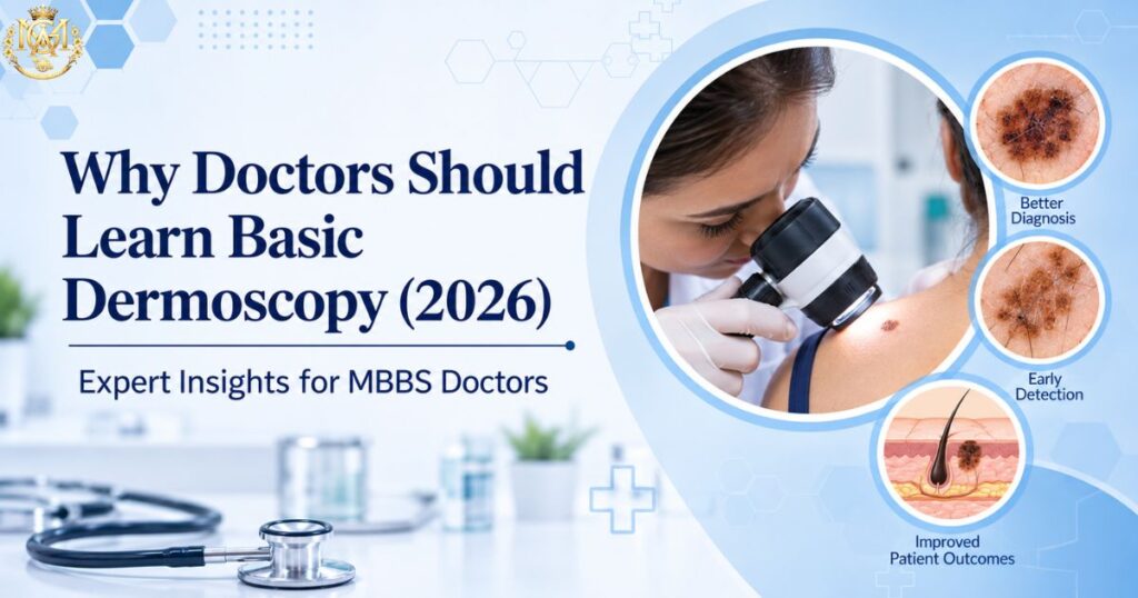
Graphic illustrating why doctors should learn basic dermoscopy, showing a physician examining a patient alongside magnified skin lesions
Learning blogs

Why Every General Physician Should Learn Basic Dermoscopy

📅 Updated September 2026 ⏰ 8 min read 🩺 General Physicians & MBBS 🌎 Dermatology Cluster — Day 8 ⚡ Quick Answer Why should general physicians learn basic dermoscopy? Dermoscopy is a non-invasive technique using a handheld magnifying device to see skin structures below the surface that aren’t visible to the naked eye. It’s rarely taught during MBBS, yet it meaningfully improves diagnostic confidence in exactly the situations GPs face daily — distinguishing fungal infections from eczema or psoriasis, and recognising when a pigmented lesion looks atypical enough to warrant urgent referral. It doesn’t replace clinical judgement or specialist referral — it adds a second layer of information before you decide to treat or refer. Ask most MBBS graduates what a dermatoscope is, and you’ll often get a vague answer — “something dermatologists use to look at skin.” That vagueness reflects a real gap: structured dermoscopy training for general physicians remains rare, and most GPs go through an entire career without direct exposure to the skill, despite skin complaints being among the most common reasons patients walk into a general OPD. TThis is worth addressing directly. Dermoscopy isn’t an obscure specialist tool — it’s a practical skill that sharpens the differential diagnosis decisions GPs make constantly. Skin presentations are among the most frequent reasons patients seek a general consultation. Yet the diagnostic toolkit most GPs bring to that consultation hasn’t changed much beyond visual inspection and history-taking. Dermoscopy fills a genuine gap in that toolkit, without requiring a full dermatology residency to gain real value from it.. This guide covers what dermoscopy actually is, what it can help a non-specialist recognise, where it fits into common differential diagnosis dilemmas, its role in early skin cancer awareness, and what structured learning actually looks like. What Is Dermoscopy, and Why Do So Few General Physicians Get Trained In It?y Dermoscopy uses a handheld device called a dermatoscope. It combines magnification with a light source, either polarised or in direct contact with the skin using an interface fluid. This lets doctors visualise structures beneath the skin’s surface that aren’t visible to the naked eye. This includes pigment patterns, vascular arrangements, and follicular openings that carry real diagnostic information. The tool itself isn’t complex or expensive to use, but three factors keep it largely confined to dermatology practice. First, it simply isn’t part of the standard MBBS curriculum at most institutions, so exposure typically only happens during dermatology postgraduate training. Second, interpreting what you see through a dermatoscope takes structured learning — pattern recognition here isn’t intuitive the way basic clinical examination is. Third, there’s a common misconception that dermoscopy is purely a cancer-screening tool for specialists, which undersells its everyday value in routine differential diagnosis. This is somewhat unusual compared to other diagnostic aids in medicine. A stethoscope, an otoscope, or a basic ophthalmoscope are all standard equipment GPs learn to use during MBBS. Each of these tools has its own learning curve too. Dermoscopy sits in a strange gap — genuinely useful, not prohibitively difficult to learn at a basic level. Yet core curricula rarely introduce it. That gap is exactly what makes it worth actively seeking out rather than waiting to encounter it by chance What Dermoscopy Can Help a Non-Specialist Recognise Even at a basic, non-expert level, dermoscopy adds genuinely useful information to a standard clinical exam. Vascular Pattern Recognition Distinguishing the type and arrangement of blood vessels visible in a lesion — a detail invisible to the naked eye but often characteristic of specific conditions. Scale & Surface Structure Identifying scale patterns, follicular plugging, and comedo-like openings that help differentiate inflammatory from infective skin conditions. Pigment Network Assessment Seeing whether a pigmented lesion’s structure looks regular and symmetric, or irregular enough to warrant a closer specialist look. Hair & Follicular Changes Recognising characteristic hair shaft changes that support a fungal diagnosis in hair-bearing skin, alongside standard KOH testing. The value here isn’t that dermoscopy replaces clinical judgement — it’s that it adds a second, objective layer of information before you decide whether to treat, watch, or refer. That’s a meaningfully different position than relying on morphology and history alone. Dermoscopy in Differential Diagnosis: Tinea, Eczema, Psoriasis This is where dermoscopy earns its keep in daily general practice. Our earlier guide on eczema vs psoriasis vs fungal rash covers the clinical differentiators between these three commonly confused conditions — dermoscopy adds a further layer of objective evidence on top of that clinical picture. Condition What Dermoscopy Adds Tinea (Fungal) Characteristic hair shaft changes in affected hair-bearing skin, supporting the diagnosis alongside KOH testing Psoriasis A fairly regular, evenly distributed vascular pattern against a light background, with diffuse white scale Eczema Less specific findings — often patchy vessel distribution with a yellowish scale or serous crust pattern, distinct from psoriasis’s regularity This is particularly valuable in exactly the scenario our differential diagnosis guide flags as the trickiest: steroid-modified presentations. In these presentations, inappropriate steroid cream use has altered a fungal infection that no longer shows classic clinical features. Combining dermoscopy findings with a KOH scraping adds meaningfully more diagnostic confidence in these atypical cases than clinical morphology alone.” Early Skin Cancer Screening: What General Physicians Should Know About Dermoscopy Dermoscopy is well established in dermatology as improving diagnostic accuracy for pigmented lesion assessment compared to naked-eye examination alone — a finding echoed in a systematic review of dermoscopy use for melanoma detection and triage in primary care settings. For general physicians, the relevant takeaway isn’t to become a diagnostician for skin cancer — it’s to develop enough pattern awareness to recognise when a pigmented lesion looks atypical and needs prompt specialist referral, rather than a “watch and wait” approach. Broad awareness points worth building familiarity with include asymmetry in a lesion’s shape and irregular or poorly defined borders. Others include colour variation within a single lesion, and any lesion changing in size, shape, or appearance over time. None of these features alone confirm malignancy. A non-specialist shouldn’t rely on any

Surgeons in an operating room representing MS surgical specialization training and lifestyle
Learning blogs

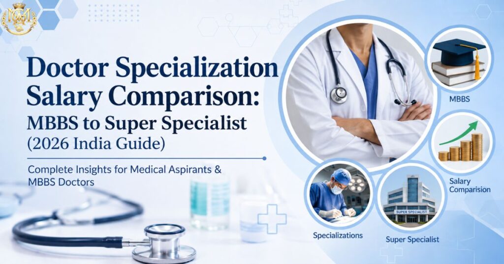
Doctor Specialization Salary Comparison: MBBS to Super Specialist (2026 India Guide

📅 Updated September 2026 ⏰ 11 min read 🎓 MBBS to Super Specialist 🌎 Specialization × Salary Cluster ⚡ Quick Answer How does doctor salary in India change from MBBS to super specialist? This doctor specialization salary comparison shows how pay scales in stages by qualification level, not just experience. An MBBS fresher typically earns ₹40,000–₹90,000 a month. An MD/MS consultant moves into the ₹1 lakh–₹3.3 lakh range depending on seniority. An established DM/MCh super specialist can earn ₹4.2 lakh–₹16.7 lakh a month, with senior national-level experts reaching ₹6.7 lakh–₹25 lakh. Every figure below is pulled directly from the same reference data used across our complete doctor salary guide. That keeps the numbers here fully consistent with it. Why This Doctor Specialization Salary Comparison Starts With Qualification Level This doctor specialization salary comparison begins with a common question doctors ask: “which specialty pays the most?” But the more useful question is how income actually changes as you move up the qualification ladder. The honest answer is that salary in Indian medicine scales in fairly distinct stages. Those stages are MBBS, then MD or MS, then DM/MCh for those who go on to super-specialise. Each step doesn’t just add a bit more pay — it typically unlocks a meaningfully different earning band. This guide walks through the full ladder using the same reference data behind our complete doctor salary guide. Whether you’re comparing a single stage or the entire career arc, the numbers stay consistent. You won’t find contradictions anywhere else on the site. If you’re still deciding which specialty to pursue, look at this ladder first. It matters more than any single specialty’s headline number. Two doctors in very different fields — a General Medicine physician and an Orthopaedic surgeon — can earn similar incomes. That’s simply because they’re at the same qualification stage. The specialty-specific differences described in our highest paying medical specializations guide matter too. But they become far more meaningful once you’ve factored in where you sit on this broader ladder. How Doctor Salaries Scale With Qualification Level Three things drive where you sit on the salary ladder at any point. The first is your qualification level — MBBS, MD/MS, or DM/MCh. The second is your years of experience within that level. The third is your practice setting — government, private hospital, or independent practice. Qualification level sets the band you’re in. Experience moves you up within that band. Practice setting determines where within the range you actually land. What’s worth understanding upfront is that these bands overlap. A highly experienced MBBS doctor without a postgraduate degree can, in some cases, out-earn a newly qualified MD consultant. But the ceiling for further growth is structurally different. Postgraduate and super-specialist qualifications don’t just pay more on average; they unlock significantly higher ceilings as experience accumulates. It’s also worth being clear about what these figures represent. Each range reflects typical monthly pay across government postings, private hospitals, and independent or consulting-based practice. It isn’t a single fixed number. Where you land within a given range depends heavily on which setting you’re in. It also depends on your specific city and institution. Two doctors at the identical qualification stage can sit at opposite ends of the same range for exactly this reason. MBBS-Level Earnings (Internship to 10+ Years, No PG) Before any postgraduate qualification, earnings still grow meaningfully with experience. That growth just sits within a narrower band than what postgraduate training eventually unlocks. Career Stage Monthly Range Internship ₹15,000 – ₹35,000 MBBS Fresher (0–2 yr) ₹40,000 – ₹90,000 MBBS 3–5 years ₹80,000 – ₹1.5 lakh MBBS 5–10 years ₹1 lakh – ₹2 lakh MBBS Senior (10+ yr, no PG) ₹1.5 lakh – ₹2.2 lakh Internship is a training stipend, not a full salary. It reflects your status as a student completing mandatory clinical rotations rather than an independently practising doctor. Once you cross into fresher territory, most doctors take roles like Medical Officer, RMO, or Duty Doctor. These roles are stepping stones while they decide their next move. The jump from fresher to the 3–5 year mark typically reflects growing clinical confidence. In many cases, it also marks a shift from purely rotational duty roles into more defined, better-compensated positions. By the 5–10 year mark without a postgraduate qualification, earnings tend to plateau. This is precisely the point at which MD, MS, or DNB training becomes the clearest lever for continued income growth. It’s not just about clinical growth anymore. The gap between MBBS Senior (₹1.5 lakh–₹2.2 lakh) and MD Senior Consultant (₹2.1 lakh–₹3.3 lakh) illustrates this well. A postgraduate qualification doesn’t just add incremental pay — it opens an entirely different ceiling for the years that follow. MD Salary Progression (Junior to Senior Consultant) Career Stage Monthly Range MD Junior Consultant (0–3 yr) ₹1 lakh – ₹2.1 lakh MD Senior Consultant (4–10 yr) ₹2.1 lakh – ₹3.3 lakh Completing an MD moves you into consultant-level roles across physician-track branches. These include General Medicine, Dermatology, Radiology, Paediatrics, Psychiatry, Pathology, and others. The jump from junior to senior consultant status typically tracks with growing diagnostic autonomy and a larger patient base. It also often means a shift from purely hospital-employed roles toward some mix of hospital and private consultation. Within this range, individual branches still vary. A Dermatology or Radiology consultant with a growing private practice may reach the top of the senior consultant band sooner. A purely hospital-employed physician in a lower-volume department may take longer. The qualification stage sets the band; the specific branch and practice model determine where within it you land. MS Salary Progression (Junior to Independent Surgeon) Career Stage Monthly Range MS Junior Surgeon (0–3 yr) ₹1.25 lakh – ₹2.5 lakh MS Independent Surgeon (4–10 yr) ₹2.5 lakh – ₹6 lakh+ Surgical branches include General Surgery, Orthopaedics, ENT, Obstetrics & Gynaecology, and Ophthalmology. These branches show a notably wider top-end range at the independent surgeon stage. This reflects how surgical income often combines a base hospital or consulting role with procedure-based private practice. Operative

Scroll to Top