
Dermoscopy for General Physicians: Skin Diagnosis, Differential Diagnosis & When to Refer
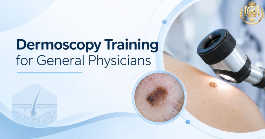
📅 Updated September 2026 ⏰ 8 min read 🩺 General Physicians & MBBS 🌎 Dermatology Cluster — Day 8 ⚡ Quick Answer Why should general physicians learn basic dermoscopy? It’s rarely taught during MBBS. Yet it meaningfully improves diagnostic confidence in exactly the situations GPs face daily — distinguishing fungal from eczema or psoriasis, and recognising when a pigmented lesion needs urgent referral. It doesn’t replace clinical judgement or specialist referral — it adds a second layer of information before you decide to treat or refer. Ask most MBBS graduates what a dermatoscope is, and you’ll often get a vague answer — “something dermatologists use to look at skin.” That vagueness reflects a real gap. Dermoscopy is rarely part of the standard MBBS curriculum. Most general physicians go through an entire career without seeing it — despite skin complaints being one of the most common reasons patients walk into a general OPD.. This is worth addressing directly, because dermoscopy isn’t an obscure specialist tool — it’s a practical skill that sharpens exactly the kind of differential diagnosis decisions GPs make constantly. Skin presentations are among the most frequent reasons patients seek a general consultation. Yet the diagnostic toolkit most GPs bring to that consultation hasn’t changed much. It still relies mainly on visual inspection and history-taking.. Dermoscopy fills a genuine gap in that toolkit, without requiring a full dermatology residency to gain real value from it. This guide covers what dermoscopy is and what it helps a non-specialist recognise. It also covers its role in differential diagnosis, skin cancer awareness, and what structured learning looks like.. What Is Dermoscopy and Why It’s Underused Outside Dermatology Dermoscopy uses a handheld device called a dermatoscope. It combines magnification with a light source — either polarised or in contact with the skin via an interface fluid. This lets you visualise structures beneath the skin’s surface that aren’t visible to the naked eye.. This includes pigment patterns, vascular arrangements, and follicular openings that carry real diagnostic information. The tool itself isn’t complex or expensive to use, but three factors keep it largely confined to dermatology practice. First, it simply isn’t part of the standard MBBS curriculum at most institutions, so exposure typically only happens during dermatology postgraduate training. Second, interpreting what you see through a dermatoscope takes structured learning — pattern recognition here isn’t intuitive the way basic clinical examination is.Third, many doctors assume dermoscopy is purely a cancer-screening tool for specialists. That misconception undersells its everyday value in routine differential diagnosis. This is somewhat unusual compared to other diagnostic aids in medicine. A stethoscope, an otoscope, or a basic ophthalmoscope are all standard equipment GPs are trained to use during MBBS, despite requiring their own learning curve. Dermoscopy sits in a strange gap — genuinely useful, not prohibitively difficult to learn at a basic level, yet rarely introduced as part of core training. That gap is exactly what makes it worth actively seeking out rather than waiting to encounter it by chance. What Dermoscopy Can Help a Non-Specialist Recognise Even at a basic, non-expert level, dermoscopy adds genuinely useful information to a standard clinical exam. Vascular Pattern Recognition Distinguishing the type and arrangement of blood vessels visible in a lesion — a detail invisible to the naked eye but often characteristic of specific conditions. Scale & Surface Structure Identifying scale patterns, follicular plugging, and comedo-like openings that help differentiate inflammatory from infective skin conditions. Pigment Network Assessment Seeing whether a pigmented lesion’s structure looks regular and symmetric, or irregular enough to warrant a closer specialist look. Hair & Follicular Changes Recognising characteristic hair shaft changes that support a fungal diagnosis in hair-bearing skin, alongside standard KOH testing. The value here isn’t that dermoscopy replaces clinical judgement — it’s that it adds a second, objective layer of information before you decide whether to treat, watch, or refer. That’s a meaningfully different position than relying on morphology and history alone. Dermoscopy in Differential Diagnosis: Tinea, Eczema, Psoriasis This is where dermoscopy earns its keep in daily general practice. Our earlier guide on eczema vs psoriasis vs fungal rash covers the clinical differentiators between these three commonly confused conditions — dermoscopy adds a further layer of objective evidence on top of that clinical picture. Condition What Dermoscopy Adds Tinea (Fungal) Characteristic hair shaft changes in affected hair-bearing skin, supporting the diagnosis alongside KOH testing Psoriasis A fairly regular, evenly distributed vascular pattern against a light background, with diffuse white scale Eczema Less specific findings — often patchy vessel distribution with a yellowish scale or serous crust pattern, distinct from psoriasis’s regularity This is particularly valuable in exactly the scenario our differential diagnosis guide flags as the trickiest: steroid-modified presentations, where a fungal infection has been altered by inappropriate steroid cream use and no longer shows classic clinical features. In these atypical cases, combining dermoscopy findings with a KOH scraping provides meaningfully more diagnostic confidence than relying on clinical morphology alone. Dermoscopy for Early Skin Cancer Screening Awareness Dermoscopy is well established in dermatology as improving diagnostic accuracy for pigmented lesion assessment compared to naked-eye examination alone. For general physicians, the relevant takeaway isn’t to become a diagnostician for skin cancer — it’s to develop enough pattern awareness to recognise when a pigmented lesion looks atypical and needs prompt specialist referral, rather than a “watch and wait” approach. Broad awareness points worth building familiarity with include asymmetry in a lesion’s shape, irregular or poorly defined borders, variation in colour within a single lesion, and any lesion that’s changing in size, shape, or appearance over time. None of these features alone confirm malignancy, and none of them should be used as a standalone diagnostic checklist by a non-specialist — they’re a screening lens for deciding who needs a dermatologist’s assessment sooner rather than later. ⚠️ Dermoscopy at the GP level is a triage and referral tool, not a diagnostic endpoint. Any lesion that looks atypical on dermoscopic examination still requires dermatology assessment and, where indicated, biopsy —