FELLOWSHIP IN NEURORADIOLOGY
Discover the world of neuroradiology fellowship with our practice-focused postgraduate diploma program. Ideal for medical professionals looking to enhance their diagnostic skills, our course offers clinical training and expert mentorship.
Batch starts on
Jan to Dec/ May to June
Course Duration
12 Months
Multimodal Program
Hybrid Mode & Clinical Attachment
Flexible payment
financing options available
Sample Certificate For Course
FELLOWSHIP IN NEURORADIOLOGY
The Neuroradiology Fellowship at MGA Medical Global Academy is an entire training program that provides radiologists with an opportunity to specialize in neuroimaging. The fellowship program is an integration of both academic and clinical training, which allows fellows to acquire the knowledge of more complex imaging modalities such as MRI, CT and PET scans. Participants will be involved in case discussions, multidisciplinary conferences and research opportunities, and will improve their diagnostic skills and knowledge of neurological disorders. Upon completion of the fellowship, the graduates will be equipped to play a role in the clinical practice as well as the academic research of the neuroradiology practice and become the leaders of this rapidly changing area.
What you'll learn
Fundamentals of Neuroradiology

Advanced MRI Techniques
Neurovascular Imaging
Neurodegenerative Disorders

Tumor Imaging
Pediatric Neuroradiology
Key Features
- Attending /Non-Attending Classes
- Clinical Training in Nearest Hospital/Clinics.
- Free Study Material with recorded lectures
- Best Faculties (Super Specialists) of India
5. Library Access & Question & Answer Session
6. NAAC A+ University
7. 1 Year of Mentorship
8. Updated Curriculum
Course Curriculum
- Neuroanatomy of the brain, spine, and head & neck
- Principles of CT, MRI, angiography, and advanced neuroimaging
- Imaging physics, safety, and protocols
- Contrast agents: techniques, safety standards, and 2025 guidelines
- Introduction to structured reporting in neuroradiology
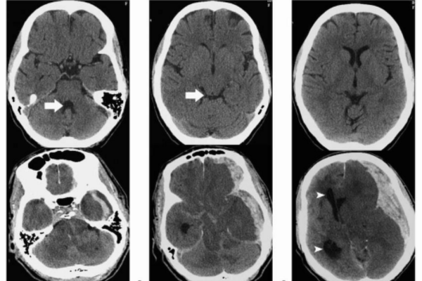
- Stroke imaging: CT perfusion, MRI diffusion, penumbra assessment
- Hemorrhage patterns & neurovascular emergencies
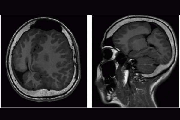
- Brain tumors—classification, grading, and multiparametric MRI
- Infection and inflammation: meningitis, encephalitis, autoimmune disorders
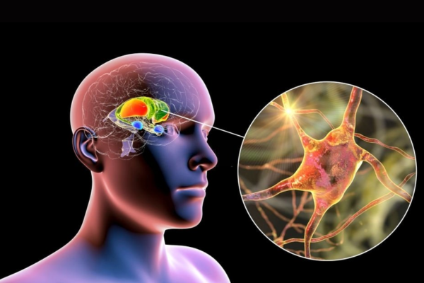
- Neurodegenerative diseases: dementia protocols, volumetry, PET correlation
- fMRI (BOLD imaging), DTI/tractography, MR spectroscopy (MRS)
- Perfusion-weighted imaging (PWI): DSC, DCE, ASL
- Spinal cord tumors and neoplasms
- Functional connectomics and emerging AI-assisted MRI analytics
- Degenerative spine disease and spinal stenosis
- Trauma imaging & spinal injury classification
- Spinal tumors and metastatic disease
- Inflammatory and demyelinating diseases
- Postoperative spine imaging and hardware evaluation
- Sinonasal disease, orbital pathology, skull base lesions
- Head & neck tumors: imaging, staging, and postoperative evaluation
- Temporal bone imaging and otologic disorders
- Dental and maxillofacial imaging principles
- Vascular lesions of the head & neck
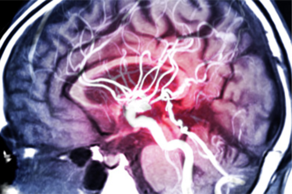
- CTA, MRA, DSA interpretation
- Aneurysms, AVMs, vasculitis, venous thrombosis
- Stroke triage: 2025 thrombectomy selection criteria
- Carotid and vertebral artery imaging
- Perfusion and collateral assessment techniques
- Congenital brain malformations
- Pediatric brain tumors and metabolic diseases
- Pediatric spine and trauma imaging
- Neonatal imaging: ultrasound, MRI, hypoxic-ischemic injury
- Radiation safety in children
- Image-guided pain management (spine injections, nerve blocks)
- Lumbar puncture, myelography, cisternography
- Fundamentals of endovascular procedures
- Emergency interventions and workflow simulation
- Post-treatment imaging evaluation
- AI-based image analysis and workflow integration (2025 standards)
- Quantitative imaging and radiomics
- Neuro-oncology imaging biomarkers
- Writing scientific papers and conducting clinical research
- Ethics, documentation, and global data privacy compliance
- Rapid stroke triage imaging protocols (2025 updated guidelines)
- Trauma: brain, spine, and maxillofacial emergency patterns
- Acute infections, encephalitis, and rapid deterioration cases
- Workflow optimization for emergency departments
- Radiology reporting in critical care environments
- MRI protocols for epilepsy (3T & 7T MRI trends)
- Localization imaging: hippocampal sclerosis, cortical dysplasia
- Pre-surgical evaluation using fMRI, PET, SPECT, MEG correlation
- Functional connectivity and seizure network mapping
- Postoperative evaluation of epilepsy surgery
- Imaging biomarkers for tumor progression vs. pseudoprogression
- Radiomics in brain tumor classification
- Advanced perfusion and spectroscopy for gliomas
- Post-treatment imaging: radiation necrosis, immunotherapy effects
- Tumor response imaging using RANO 2025 criteria
- MRI criteria for MS (2025 McDonald updates)
- Differentiating MS from NMOSD, MOGAD, ADEM
- Spinal cord involvement assessment
- Quantitative lesion load analysis and volumetric tools
- Monitoring disease activity and therapy response
- Optic nerve and orbital imaging
- Visual pathway disorders
- Vascular compression syndromes
- Pituitary and skull base lesions affecting vision
- Ocular trauma and emergency imaging
- Brain connectivity networks and default mode network assessment
- Imaging of psychiatric disorders (2025 imaging markers)
- Dementia differentiation: Alzheimer’s, FTD, DLB, vascular
- Quantitative brain volumetry and cortical thickness analysis
- PET-MRI hybrid imaging interpretation
- MRI neurography protocols
- Brachial plexus and lumbosacral plexus imaging
- Peripheral nerve tumors and entrapment syndromes
- Image-guided therapeutic procedures
- Neuropathic pain imaging patterns
- Dual-energy CT for neuro applications
- Ultra-high-resolution CT for skull base and temporal bone
- CT perfusion optimization and artifact reduction
- Radiation dose–reduction strategies (2025 technologies)
- CT angiography pitfalls and advanced reconstructions
- Bedside CT/MRI protocols and portable imaging
- Monitoring cerebral edema, herniation, and intracranial pressure
- Imaging in ECMO, ventilated, and unstable patients
- Post-cardiac arrest encephalopathy imaging
- Critical care decision-making: radiologist’s role
- CNS tuberculosis, neurocysticercosis, and parasitic infections
- Fungal infections and immunocompromised brain imaging
- Emerging 2025 tropical neuroinfections
- Differentiating infectious from inflammatory lesions
- Head & neck infectious complications
- Imaging in hepatic, uremic, and mitochondrial disorders
- Toxic exposures: alcohol, drugs, environmental neurotoxins
- Metabolic disorders in neonates and adults
- Advanced DWI/DTI for metabolic patterns
- Radiologic–clinical correlation strategies
- Preoperative mapping for endoscopic skull base surgery
- Sinonasal tumor staging with AI-assisted planning
- Cranial nerve pathway mapping
- Orbit–sinus–skull base anatomical variants
- Virtual navigation imaging for surgeons
Our Accrediation Partners

Fill Form Our Team Will Contact Soon
Choose medicalglobalacademy as your trusted partner for medical education and elevate your expertise to new heights.
Program Highlights
- Duration: 12 Months
- Type: On-line + Clinical Training Hybrid.
- Focus Areas: Brain, Spine & Head-Neck Imaging
- Mentorship: Expert Neuroradiologists & Imaging Specialists
- Accreditation: CPD UK Certified
- Learning Mode: Flexible, aimed at working professionals.
Program Structure
Phase I: Academic and Theoretical training (Online)
This step is aimed at establishing a solid background in neuroradiology principles and interpretation of images.
Curriculum Focus:
- Correlations between neuroanatomy and imaging.
- MRI & CT imaging techniques
- Physics of imaging and optimization of protocols.
- Structured reporting and diagnostic approach
- Basics of sophisticated neuroimaging.
Learning Components:
- Live interactive classes with faculty of experts.
- Taped lectures to allow flexible access.
- Case-based discussions and clinical reviews.
- Organized learning material and reference materials.
Phase II: Clinical & Imaging Training
This stage focuses on practical training and field-work exposure to diagnosis.
Clinical Training Includes:
- Reading of actual neuroimaging cases.
- Monitored reporting and diagnostic processes.
- High-volume radiology department exposure.
- Involvement in multidisciplinary discussions.
Core Clinical Areas:
- Neuro-oncology imaging and brain tumors.
- Stroke and vascular imaging (MR angiography)
- The neurodegenerative and demyelinating disorders.
- Brain and spinal injuries that are traumatic.
- Pediatric neuroradiology
- Head & neck imaging
Advanced Imaging Exposure
- Students are provided with the field experience in the latest diagnostic methods:
- Functional MRI (fMRI)
- Diffusion Tensor Imaging (DTI)
- MR Spectroscopy
- Perfusion imaging
- Post-operative and spinal imaging examination.
Clinical Training Environment
- The training will take place in the most prominent radiology departments and neuroimaging centers.
- Experience with varied and multifaceted case profiles.
- Liaison with neurosurgeons, neurologists, and oncologists.
- Pay attention to accuracy of reporting, accuracy of reporting, and integration into clinical.
Assessment & Certification
- Evaluation at the end of training.
- MCQ-based evaluation
- Fellowship certification accredited by CPD UK
- Identification was in line with international imaging standards.
Eligibility Criteria
- Radiologists
- Neurologists
- MBBS graduates
- Physicians who are interested in the specialization of neuroimaging.
Career Advancement
- Participants will be able to:
- Independently interpret complicated neuroimaging studies.
- Utilize the latest imaging modalities in clinical practice.
- Enhance the quality of diagnosis and reporting.
- Establish a good career in radiology in hospitals, universities, or diagnostics.
Our top courses
fetal medicine fellowship
fellowship in obstetrics & gynaecology
fellowship in musculoskeletal ultrasound
fellowship in Clinical Cardiology
dermatology fellowship
fellowship in clinical neurology
fellowship in oncology
robotic surgery fellowship
fellowship in internal medicine
Frequently Asked Questions
To apply, visit the course page of your interest and click on the ‘Apply Now’ button. Follow the instructions to complete your application. You may need to submit relevant documents and a resume.
Yes, all our courses are accredited by recognized medical and educational institutions. Details of accreditation can be found on each course page.
Yes, we offer a variety of online courses to provide flexibility for our students. Check the course descriptions for information on whether a course is available online.
You can contact our support team via email at support@medicalglobalacademy.com or call us at [+919310027474]. Our support team is available Monday to Friday from 9 AM to 6 PM.