
MGA Clinical Education Series · 8 min read
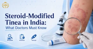
A patient walks into your OPD with a widespread, pale, mildly itchy skin rash on the trunk and groin. He has seen a pharmacist, used a cream, felt better for a week — and now it is back, larger and harder to define. This scenario plays out in dermatology OPDs and general physician clinics across India every single day. The culprit, in many of these cases, is not a new or unusual pathogen. It is an old fungal infection that has been allowed to change its face — because of a tube of OTC cream.
This article explains what steroid-modified tinea is, why India is at the centre of a documented dermatology crisis, how these altered infections present clinically, why they are harder to clear,
and what the published evidence says every treating doctor should do differently.
🔍 Quick Reference
Condition: Steroid-modified tinea / Tinea incognito
Cause: OTC topical steroid (or steroid–antifungal–antibiotic triple combination) applied to an undiagnosed dermatophyte infection
Effect: Atypical clinical appearance, missed diagnosis, wider spread, treatment resistance
Key Indian literature: IJDVL — Verma, Panda et al. 2021 (3-part series); IADVL–ITART Consensus 2020; Dutta, Rasul & Boro, IJDVL 2017
Doctor’s role: Clinical recognition, KOH confirmation, patient education, pharmacist communication
🔬 What Is Steroid-Modified Tinea (Tinea Incognito)?
Tinea incognito — now more accurately called steroid-modified tinea in Indian clinical literature — refers to a dermatophyte infection whose typical clinical appearance has been altered by the application of a topical corticosteroid (or a calcineurin inhibitor). The term was first reported in 1968 by British dermatologists Dr Adrian Ive and Dr Ronnie Marks, who described patients with fungal infections masked by topical steroid use presenting as seborrheic dermatitis, eczema, or psoriasis-like eruptions.
The mechanism is well understood: topical corticosteroids suppress local skin immunity — specifically, they downregulate antigen-presenting cell function, reduce IL-12 and Th1 activation, and shift the immune response toward a Th2 profile. The dermatophyte encounters a local immune environment that has been instructed to stand down. The inflammation that normally produces the classic raised, scaly, well-defined ring fades. The infection spreads laterally and sometimes deeper, but looks far less angry than it should. As published in Journal of Clinical Medicine (Zacharopoulou et al., 2024, PMC11172699), the more topical steroid that is applied, the more extensive and unrecognisable the fungal infection becomes.
A critical nuance noted by Verma SB and colleagues in IJDVL (2021, doi: 10.25259/IJDVL_301_20) is that in the Indian epidemic, a strict “tinea incognito” — where the infection is completely unrecognisable — is less common than steroid-modified tinea, where an active border is often still visible despite morphological distortion. The IJDVL authors argue that “steroid-modified tinea” is therefore the more accurate term for the majority of Indian cases.
🏥 How an OTC Cream Habit Became a National Dermatology Concern
India’s climate — high humidity, high ambient temperature across much of the country, and monsoon seasons that last months — creates conditions strongly favourable to dermatophyte growth and spread. Tinea corporis, tinea cruris, and tinea faciei are among the most common superficial infections seen in both urban and rural OPDs. In this environment, an itchy circular rash is a predictably common presentation.
The problem is what happens next. Fixed-dose combination (FDC) creams containing a potent topical corticosteroid (typically clobetasol propionate or betamethasone dipropionate), an antifungal agent, and an antibacterial agent are widely available over the counter in India. Research published in the Indian Journal of Dermatology, Venereology and Leprology (Dutta, Rasul & Boro, IJDVL 2017) found that pharmacists were responsible for recommending these combination creams in the majority of tinea incognito cases — 78% of patients in that study were using products recommended by a pharmacist. Patients used these preparations for durations ranging from six weeks to twelve years without medical supervision.
Why do patients keep using them? The steroid component produces rapid, visible relief within a few days. Itch decreases, redness fades. Patients interpret this as a cure. When the cream is stopped, the itch returns worse than before — because the fungus has been spreading unchecked beneath a suppressed immune response. The patient reaches for the tube again. The cycle repeats.
A landmark 2021 three-part IJDVL series by Verma SB, Panda S, Nenoff P and colleagues described the situation as an “unprecedented epidemic-like scenario” of dermatophytosis in India, attributing the crisis directly to indiscriminate use of FDC corticosteroid creams and the emergence of a novel, partially terbinafine-resistant species — Trichophyton indotineae (formerly classified as T. mentagrophytes ITS genotype VIII) — which the WHO Fungal Priority Pathogens List (2022) has since acknowledged as a global concern.
📊 What the Published Research Documents
• The prevalence of superficial dermatophytosis in different regions of India ranges from 36.6% to 78.4% — a range described by experts as constituting a national epidemic (topical antifungal-corticosteroid FDC review, Indian Journal of Pharmacology).
• A cost-of-illness analysis published in Clinical and Experimental Dermatology (Jagadeesan et al., 2023) found that patients using topical steroids for dermatophytosis in India incur approximately 40% higher treatment costs than steroid-naive cases.
• The IADVL Task Force Against Recalcitrant Tinea (ITART) Consensus 2020 noted a six- to seven-year pattern of unprecedented increase in recurrent, chronic, and steroid-modified dermatophytosis involving glabrous skin.
• A study from Western Uttar Pradesh (Thakur et al., Journal of Dermatology and Cosmetology, 2018) conducted across 100 steroid-modified tinea cases identified lack of qualified dermatologists in rural areas as a compounding factor — a gap that directly increases irrational corticosteroid use.
👁️ Clinical Recognition: How Steroid-Modified Tinea Presents Differently
The diagnostic challenge is that steroid-modified tinea no longer looks like textbook ringworm. The classic well-defined, raised, scaly, erythematous ring with central clearing is modified or absent. Recognising the altered patterns is the first clinical skill every doctor managing skin conditions in India needs to develop.
| Feature | Classic Tinea (Untreated) | Steroid-Modified Tinea |
|---|---|---|
| Border | Well-defined, raised, scaly | Blurred, less elevated, less scaly |
| Central clearing | Present (classic ring shape) | Often absent — study data shows 94.1% of steroid-modified cases lack central clearing (Zenodo, 2024) |
| Colour | Erythematous (red) | Pinkish, flesh-coloured, or pale — erythema present in approximately 54% of steroid-modified cases |
| Lesion extent | Localised, well-contained | Large, diffuse, sometimes geographic or bizarre-shaped; multiple sites |
| Associated features | Scaling, mild itch | Pustular borders, secondary bacterial infection, skin atrophy, striae, telangiectasia (with prolonged steroid use) |
| What it mimics | Ringworm (recognisable) | Nummular eczema, psoriasis, seborrheic dermatitis, rosacea, granuloma annulare, lupus-like eruption |
| History clue | No prior cream use | Pharmacist/OTC cream use, temporary relief, recurrence after stopping cream |
The IJDVL landmark 2021 series (Verma et al.) also describes specific morphological variants now commonly seen in Indian OPDs: tinea pseudoimbricata (concentric rings resembling the Southeast Asian tinea imbricata pattern), tinea faciei with a rosacea-like appearance, and erythrodermic presentations extending across large body surface areas. In the IJDVL clinico-epidemiological study (Dutta et al., 2017), 70 of 100 cases of tinea incognito had two or more anatomical areas involved simultaneously — most commonly tinea faciei combined with tinea corporis and tinea cruris.
A practical clinical clue cited in the Journal of Clinical Medicine review (Zacharopoulou et al., 2024): in steroid-modified cases complicated by secondary bacterial infection, hairs can be pulled from affected follicles without pain — a finding that helps distinguish tinea with secondary infection from a primary bacterial folliculitis.
⚠️ Why Steroid-Modified Tinea Is Harder to Treat Than Typical Tinea
Three distinct factors make steroid-modified tinea significantly more difficult to clear than a straightforward, untreated tinea infection:
1 — Delayed diagnosis extends fungal spread
The atypical appearance means the condition is frequently misdiagnosed as eczema, seborrheic dermatitis, or psoriasis. Steroid treatment for these presumed diagnoses then worsens the underlying fungal infection further. By the time a correct diagnosis is reached, the infection has often spread across multiple body sites, involves hair follicles, and has invaded deeper skin layers — all of which demand longer, more aggressive antifungal courses.
2 — Antifungal resistance
The widespread and prolonged use of sub-therapeutic antifungal agents (present in OTC combination creams) has driven the selection of resistant dermatophyte strains in India. Research from Delhi (Singh et al., Mycoses, 2018) documented high terbinafine resistance rates in Trichophyton interdigitale isolates harbouring mutations in the squalene epoxidase gene. PGI Chandigarh studies (Rudramurthy et al., Antimicrobial Agents and Chemother, 2018) similarly confirmed that mutations in the ERG1 gene confer 40–80-fold reductions in terbinafine susceptibility in the Indian epidemic strains. The ITART consensus (2020) notes a notable decrease in clinical responsiveness to antifungals given in conventional doses and durations.
3 — Cycle of recurrence
Even when patients begin appropriate antifungal therapy, incomplete treatment is a recurring problem. Symptoms improve before the fungal load is fully cleared. The patient stops the antifungal. Residual organisms re-establish the infection. The IADVL ITART consensus (2020) specifically recommends that topical antifungals should be continued for two weeks beyond clinical resolution — a threshold very few patients without explicit physician instruction would reach. Additionally, patients with atopic dermatitis or other conditions requiring immunosuppressive treatment face elevated risk of recurrent infection from an impaired epidermal barrier (Zacharopoulou et al., J Clin Med, 2024).
🩺 The Doctor’s Role in Reversing the Trend
A clinically important observation from the IJDVL study (Dutta et al., 2017): there were no cases of tinea incognito caused by a dermatologist in that cohort. Every confirmed case of steroid-modified tinea in that study originated from pharmacist recommendation, self-treatment, or advice from family and friends. The doctor’s consultation — with clinical examination and, where indicated, KOH preparation — is the intervention that breaks the cycle. The doctor’s role here is both diagnostic and educational.
Take a detailed cream history
Ask specifically about OTC cream use, combination creams, duration of use, and whether symptoms initially improved then returned. Patients often do not volunteer this information unless asked. A history of temporary relief followed by a larger, more widespread rash is a strong clinical clue.
Maintain a high index of suspicion
The ITART consensus and the IJDVL series both emphasise that diagnosing tinea incognito requires a high clinical index of suspicion. In any itchy, poorly defined, widespread, pale, or atypical rash in a patient with a prior history of OTC cream use, tinea should remain on the differential — even when the presentation does not look like ringworm.
Confirm with KOH preparation where accessible
A 10% KOH preparation of skin scrapings from the lesion border (after discontinuing the topical steroid if possible) is a quick, accessible, and cost-effective confirmatory test. Scrapings should be collected from the periphery of the plaque and kept dry to prevent bacterial contamination, as specified in the ECTODERM India expert consensus (BMC Dermatology, 2018). Positive KOH showing hyphal filaments confirms a dermatophyte infection regardless of the altered clinical appearance.
Educate the patient clearly
Research shows that many patients using steroid-containing combination creams did not know they contained a potent corticosteroid or could not distinguish an antifungal from a steroid cream (Persson, Gerena-Maldonado & Woolhiser, 2025). Patient education — in the patient’s own language — about why the OTC cream felt like it worked and why it has made the infection worse is a critical part of the consultation. The IADVL’s “No Steroids for Fungal Infections” campaign provides a public health framework doctors can reference and reinforce.
Advise on hygiene measures
The dermatophyte is transmissible. Patients should be advised to avoid sharing towels, bed linen, and clothing; to wash clothing in hot water where possible; to wear loose cotton garments over affected areas; and to check close contacts and household members for similar skin changes. These measures, recommended in the ITART consensus and the IJDVL series, reduce reinfection from environmental and human sources.
📚 What the Published Literature Shows
The Indian dermatology literature on this topic is substantial, peer-reviewed, and directly practice-relevant. The key studies and consensus statements every doctor managing tinea in India should be aware of:
| Study / Publication | Journal | Key finding relevant to Indian GPs |
|---|---|---|
| Verma, Panda et al. (2021) — 3-part series | IJDVL (87:154–175, 326–332, 468–482) | Comprehensive documentation of the Indian dermatophytosis epidemic: epidemiology, diagnosis, antifungal resistance, and treatment. Establishes FDC steroid creams as the primary driver of modified tinea. |
| IADVL–ITART Consensus (Rengasamy et al., 2020) | Indian Dermatology Online Journal (11:4) | 17-expert Delphi consensus. Topical corticosteroid use — alone or in combination — strongly discouraged. Recommends continuing antifungals two weeks beyond clinical resolution. |
| Dutta, Rasul & Boro (2017) | IJDVL (83:326–331) | Clinico-epidemiological study of 100 tinea incognito cases: 78% traced to pharmacist recommendation. Zero cases were caused by a dermatologist’s prescription. Triple combinations (fluocinolone + antifungal + antibacterial) most commonly used. |
| Jagadeesan et al. (2023) | Clinical and Experimental Dermatology (48:909–912) | Cost-of-illness analysis: steroid-modified dermatophytosis patients spend approximately 40% more on treatment than steroid-naive cases, reflecting longer and more complex management. |
| Thakur et al. (2018) | Journal of Dermatology and Cosmetology (2:64–69) | 100-case study from Western UP: lack of qualified dermatologists in rural areas as a structural factor compounding irrational steroid use. |
| Zacharopoulou et al. (2024) | Journal of Clinical Medicine (13:3267, PMC11172699) | Comprehensive review of tinea incognito pathogenesis, clinical features, diagnosis, and management. Useful global reference with India-specific citations; KOH, dermoscopy, and culture guidance. |
❓ Frequently Asked Questions
Is steroid-modified tinea contagious?
Yes. The underlying dermatophyte infection remains contagious throughout. Because steroid-modified cases tend to be more widespread and prolonged, they may actually represent a greater transmission risk than a contained, properly treated tinea infection. Household contacts and partners should be examined, particularly if any are experiencing similar skin changes.
Can a KOH test be done if the patient is currently using a combination cream?
Yes, but ideally the topical steroid component should be discontinued for a few days before scraping to improve fungal element visibility. Scrapings should be taken from the active border, not the central area of the lesion, which may be sterile in modified tinea. The ECTODERM India consensus (BMC Dermatology, 2018) recommends collecting samples from the lesion periphery and keeping them dry before microscopy.
What is Trichophyton indotineae and is it different from regular tinea?
Trichophyton indotineae (formally delineated as a novel species from the T. mentagrophytes complex in 2020) is a dermatophyte that has spread in India — and now internationally — and is characterised by frequent resistance to terbinafine. Its clinical presentation largely resembles other tinea infections. It is clinically relevant because steroid-modified tinea cases in India are increasingly likely to involve this species, and standard terbinafine courses may be less effective. Species identification via culture or PCR is important in recalcitrant cases. The WHO Fungal Priority Pathogens List (2022) and the US CDC (MMWR, 2023) have both documented its global spread, with cases traced to India identified in Europe, the USA, and Canada.
Should combination steroid-antifungal-antibiotic creams ever be prescribed?
The IADVL ITART consensus (2020) strongly discourages the use of topical corticosteroid combinations — alone or in combination with other agents — for treating dermatophytosis. Even when a topical antifungal is present in the combination, the steroid component impairs local immunity sufficiently to allow the fungus to spread, as documented in the clinical literature. These products are not recommended for fungal infections, and the combination is particularly harmful when used for extended periods without medical supervision.
How is steroid-modified tinea different from recurrent tinea?
Recurrent tinea involves re-infection or relapse of a cleared infection, typically at the same site. Steroid-modified tinea involves a single continuous infection that has never been fully cleared, has been worsened by corticosteroid application, and presents with altered morphology. The distinction matters clinically: steroid-modified tinea requires stopping the cream, confirming the diagnosis, and often requires a longer antifungal course than a simple first-episode infection. For more on recurrent patterns, see our article on recurrent fungal infections in India.
📖 Continue Reading
Related Articles in This Series
→ Recurrent fungal infections in India: Why tinea keeps coming back
→ Dermatology after MBBS: The complete career and specialisation guide
Build the Clinical Skills to Recognise What Others Miss
Steroid-modified tinea is one of dozens of clinical scenarios where pattern recognition — built through structured dermatology training — changes patient outcomes. MGA’s Fellowship in Dermatology equips doctors with exactly the clinical and diagnostic depth needed to manage the full spectrum of dermatological presentations in Indian practice, including atypical and steroid-modified infections.
Disclaimer: This article is intended for qualified medical professionals and is for educational and informational purposes only. It does not constitute clinical advice or a prescribing recommendation. All clinical decisions should be based on the individual patient’s presentation, local guidelines, and the treating clinician’s professional judgement. Clinical claims in this article are referenced to published peer-reviewed literature as cited. Readers are encouraged to consult the source publications directly.