
Medical Global Academy

August 19, 2026

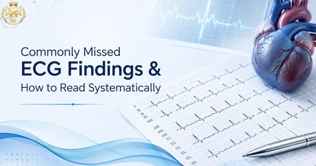
Doctor carefully reviewing an ECG to avoid common interpretation mistakes in primary care
Learning blogs

Commonly Missed ECG Findings in Primary Care and How to Systematise Your Reading

Quick Answer Most missed ECG findings in primary care aren’t missed because the doctor doesn’t know the pattern — they’re missed because time pressure replaces a systematic read with a quick glance for the obvious. The fix is a fixed reading sequence (rate, rhythm, axis, intervals, morphology, then clinical correlation) applied to every ECG, every time — plus specific familiarity with the patterns that look deceptively unremarkable: posterior MI, de Winter T waves, Wellens syndrome, early hyperkalemia, and subtle STEMI in the presence of LBBB. This article covers both the method and the specific patterns. An ECG rarely gets misread because the underlying pattern is obscure. It gets misread because a busy OPD or emergency shift compresses a two-minute systematic read into a five-second glance for the obvious — an ST elevation that jumps out, a rate that’s clearly too fast or too slow. The findings that get missed are, almost without exception, the ones that don’t announce themselves. This article covers two things together, because they solve the same problem from different angles: a systematic reading method that structurally reduces the chance of missing something, and a specific list of the patterns most often missed even by doctors reading carefully — because they look subtle, atypical, or deceptively benign at first glance. Doctors who want to build this into a formal, structured skill can explore MGA’s Certificate in ECG Interpretation, which covers systematic reading and pattern recognition in depth. 7 steps In a systematic reading method 8 patterns Most frequently missed in primary care 0 shortcuts The method works because it skips none What this article covers Why ECG misses happen even when doctors know the patterns A systematic reading method you can apply to every ECG Eight commonly missed findings — and why each one hides How to build reading speed without cutting corners Frequently asked questions Why ECG misses happen even when doctors know the patterns Ask most doctors whether they know what a de Winter T wave pattern looks like, or what Wellens syndrome represents, and many will say yes. Knowledge isn’t usually the gap. What breaks down in practice is application under pressure — a busy OPD queue, an emergency department at capacity, or simple pattern fatigue after reading dozens of unremarkable ECGs in a row. Three specific failure modes account for most misses in primary care and general emergency settings: Single-glance triage: Scanning for the one obvious abnormality — a clear ST elevation or an obviously irregular rhythm — and stopping there, rather than completing a full systematic read regardless of what’s found early. Anchoring on a normal-looking baseline: Several high-stakes patterns (posterior MI, Wellens syndrome, early hyperkalemia) present with a standard 12-lead ECG that looks largely unremarkable at first pass, because the abnormality is subtle, reciprocal, or in a lead group that’s easy to underweight. Isolated reading without clinical correlation: Reading the ECG as a standalone image rather than integrating it with the presenting complaint. A patient who was pain-free at the time of the ECG but describes classic exertional chest pain in the history needs the ECG read with that context in mind — a resting, asymptomatic ECG can look deceptively reassuring in exactly this scenario. A systematic reading method you can apply to every ECG The single highest-value habit in ECG interpretation is applying the same fixed sequence to every ECG, regardless of how obvious or unremarkable it appears at first glance. The sequence below is a standard, widely taught structure — the value isn’t in any single step being novel, it’s in never skipping one. 1. Rate Establish the ventricular rate first — both to flag obvious tachycardia or bradycardia and because rate context changes how you interpret everything that follows. 2. Rhythm Determine whether the rhythm is regular or irregular, and whether it’s sinus. This step alone catches atrial fibrillation, flutter, and most significant arrhythmias — but only if performed deliberately rather than assumed from a “normal-looking” rate. 3. Axis Assess the QRS axis. Axis deviation can point toward chamber hypertrophy, conduction abnormalities, or prior infarction — findings that are easy to skip because axis assessment feels like an extra step rather than an obviously urgent one. 4. Intervals Measure the PR interval (looking for heart block), QRS duration (looking for bundle branch block or conduction delay), and QT interval (looking for prolongation relevant to arrhythmia risk and drug interactions). Each interval answers a different clinical question — none should be skipped because another looks normal. 5. P wave and QRS morphology Examine P wave shape and consistency (atrial enlargement, ectopic atrial rhythms) and QRS morphology (bundle branch blocks, ventricular hypertrophy, pathological Q waves indicating prior infarction). 6. ST segment and T wave This is where most acute ischemic findings live — and where most subtle misses happen. Assess every lead group systematically for ST elevation, ST depression, and T wave changes, including reciprocal changes in leads that aren’t the “obvious” ones for the clinical presentation. 7. Clinical correlation Integrate the ECG findings with the presenting complaint, risk factors, and timing of symptoms. An ECG is a snapshot — a normal or near-normal ECG in a patient with a highly suggestive history does not rule out a significant cardiac event, particularly if the ECG was taken between symptomatic episodes. Build a formal, structured ECG interpretation skill MGA’s Certificate in ECG Interpretation — systematic reading, rhythm recognition, ischemia patterns View Program Eight commonly missed findings — and why each one hides Finding What It Looks Like Why It’s Missed Posterior MI ST depression and tall R waves in V1–V3 — a reciprocal, mirror-image pattern rather than direct ST elevation No ST elevation is visible on the standard 12-lead; the abnormality is a mirror pattern in anterior leads, easily read as “unremarkable” De Winter T wave pattern Upsloping ST depression with tall, symmetric T waves in the precordial leads Absence of ST elevation despite representing an acute proximal LAD occlusion equivalent — easily read as non-specific change Wellens syndrome Deeply
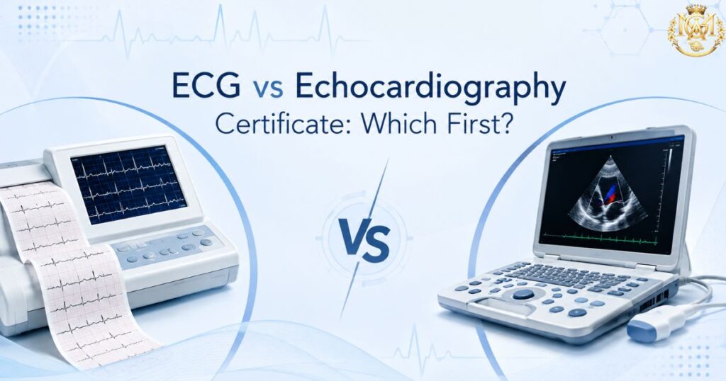
Doctor comparing clinical skills for a certificate in ECG interpretation and echocardiography
Learning blogs

Certificate in ECG Interpretation vs Certificate in Echocardiography: Which Cardiac Skill First?

Quick Answer For most MBBS doctors, general physicians, and emergency practitioners, ECG interpretation is the stronger first choice — it requires no equipment beyond what most clinics already have, applies to nearly every patient encounter involving chest pain or palpitations, and builds a diagnostic reflex you’ll use daily. Echocardiography is the better first choice specifically if you already work alongside an ultrasound machine or want to add bedside cardiac imaging to your practice. Many doctors eventually complete both — the sequencing question is really about which skill pays off faster in your specific setting. Cardiac skill-building is rarely an all-or-nothing decision. Most doctors don’t have the time or need for a full cardiology fellowship, but they do encounter chest pain, palpitations, and breathlessness often enough that a focused cardiac skill genuinely changes how confidently they manage a patient. The real question most doctors face isn’t “should I learn cardiology” — it’s “which specific skill should I build first.” MGA’s Certificate in ECG Interpretation and Certificate in Echocardiography are two distinct answers to that question. This article compares them directly — what each actually teaches, what equipment and setting each assumes, and which one to prioritise based on your current practice. If you’re still exploring the full range of cardiology course formats available after MBBS, our cardiology courses after MBBS guide covers that broader landscape; this article is specifically about sequencing these two certificates. 2 skills Compared for sequencing, not ranked Zero vs Equipment Core practical difference Daily use Both apply to routine OPD and ER work What this guide covers What the Certificate in ECG Interpretation covers What the Certificate in Echocardiography covers Direct comparison: equipment, learning curve, and daily use Who should learn ECG interpretation first Who should learn echocardiography first Can you build both skills — and in what order? Frequently asked questions What the Certificate in ECG Interpretation covers The Certificate in ECG Interpretation is built around a single, focused competency: reading a 12-lead ECG accurately and confidently, in the time pressure of a real clinical encounter — not in the unhurried setting of a textbook. Core content areas ECG fundamentals and lead placement: Cardiac electrical conduction, the relationship between the 12 leads and cardiac anatomy, and correct electrode placement — the foundation that makes every subsequent reading accurate rather than technically flawed from the start. Systematic reading method: A structured, repeatable approach to reading any ECG — rate, rhythm, axis, intervals, and morphology in a fixed sequence — designed to replace ad hoc pattern-spotting with a method that catches findings a rushed read misses. Rhythm recognition: Identifying normal sinus rhythm and the common arrhythmias encountered in general and emergency practice — atrial fibrillation, atrial flutter, supraventricular tachycardia, ventricular ectopy, and heart blocks. Ischemia and infarction patterns: Recognising ST elevation, ST depression, T wave inversion, and Q wave changes, along with the less obvious patterns that carry high clinical stakes — findings that are easy to miss under time pressure but change immediate management when caught. Clinical correlation and escalation thresholds: When an ECG finding warrants immediate referral, when it can be managed and monitored, and when it’s a benign variant — the judgment layer that separates confident interpretation from technically correct but clinically unhelpful reading. What the daily use looks like ECG interpretation requires no equipment beyond a standard ECG machine — already present in most clinics, OPDs, and emergency departments. The skill applies immediately to any patient presenting with chest pain, palpitations, breathlessness, or syncope, making it one of the most frequently used cardiac skills in general practice. View the Certificate in ECG Interpretation Focused, applied cardiac skill training · Eligible doctors View Program What the Certificate in Echocardiography covers The Certificate in Echocardiography is built around bedside cardiac ultrasound — a fundamentally different skill from ECG in that it involves image acquisition and spatial interpretation, not just waveform reading. Core content areas 2D echo fundamentals and transducer technique: Basic ultrasound physics as applied to cardiac imaging, transducer positioning, and the standard transthoracic echocardiography (TTE) windows — parasternal, apical, subcostal, and suprasternal views. Chamber assessment: Systematic evaluation of the four cardiac chambers — size, wall thickness, and gross structural assessment — building the visual vocabulary needed to recognise normal versus abnormal anatomy. Basic valve assessment: Visual assessment of valve structure and motion, and recognising overt valvular abnormalities — a foundational level of valve evaluation rather than comprehensive Doppler-based quantification. LV function and ejection fraction estimation: Visual (“eyeballing”) assessment of left ventricular contractility and a basic approach to estimating ejection fraction — one of the most clinically useful bedside echo skills, directly relevant to heart failure and acute cardiac presentations. When to refer for formal echocardiography: Recognising the boundary between what a focused certificate-level skill can answer and when a comprehensive echocardiographic study from a cardiologist or trained sonographer is required. What the daily use looks like Echocardiography requires access to an ultrasound machine — a real practical constraint that ECG interpretation does not carry. The skill is most immediately useful in settings where cardiac imaging equipment is already available: hospitals, diagnostic centres, and clinics with point-of-care ultrasound capability. View the Certificate in Echocardiography Bedside cardiac imaging skill training · Eligible doctors View Program Direct comparison: equipment, learning curve, and daily use Factor ECG Interpretation Echocardiography Equipment required Standard ECG machine — near-universal availability Ultrasound machine — not present in every clinic Skill type Waveform reading and pattern recognition Image acquisition + spatial interpretation Learning curve Faster — no hand-eye probe coordination needed Longer — image quality is skill-dependent Frequency of clinical use Very high — any chest pain/palpitation presentation High where equipment exists, low otherwise Standalone diagnostic value High — often decisive on its own for acute presentations High but frequently used alongside other findings Typical settings OPD, emergency department, rural/primary care Hospitals, diagnostic centres, imaging-equipped clinics Best first skill for General physicians, emergency doctors, rural practitioners Doctors already working alongside ultrasound equipment Course duration, fees, and specific eligibility should be verified on each

Scroll to Top