Medical Global Academy

Learning blogs

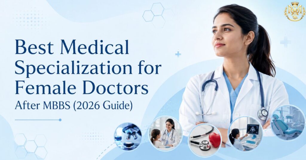
Confident female doctor representing the best medical specialization options after MBBS
Learning blogs

Best Medical Specialization for Female Doctors After MBBS (2026 Guide)

Quick Answer No medical specialty in India is restricted by gender — every branch is open to every qualified doctor. This guide covers specialties that many women doctors commonly choose for their outpatient-heavy structure and comparatively predictable schedules — Dermatology, Radiology, Pathology, Pediatrics, Obstetrics & Gynaecology, and Ophthalmology — alongside a clear-eyed look at women thriving in surgical and emergency branches too. The right specialty depends on your clinical interest, your tolerance for on-call work, and your long-term practice goals — not your gender. Search “best specialization for female doctors” and you’ll find lists that quietly assume women are choosing specialties around family planning and reduced ambition. That framing is both inaccurate and unhelpful. Women in India lead departments in neurosurgery, run Level 1 trauma centres, and head cardiac surgery units. Gender does not restrict any branch of medicine. What this guide actually addresses is a real and legitimate search pattern: many women MBBS graduates want to understand which specialties are commonly associated with more predictable outpatient schedules and lower night-call frequency — not because they are less committed to medicine, but because they are weighing genuine practical factors like call rotation, maternity leave structures, and long-term part-time practice viability alongside clinical interest, exactly as any doctor evaluating a decades-long career should. This article covers those specialties honestly, balances it with a section on women excelling in high-acuity surgical and emergency branches, and ends with a factor-based checklist so you can weigh the decision on your own terms — not a list someone else has decided is right for you. 0 Specialties restricted by gender in India 6+2 Specialty categories compared in this guide 4 factors In the decision checklist below What this guide covers Is any medical specialty restricted by gender? (No — here’s why this list exists) Specialties known for outpatient-friendly schedules Specialties with strong long-term flexibility (Dermatology, Radiology, Pathology) Women thriving in surgical & emergency branches — don’t rule these out Factors to weigh: call frequency, maternity-friendly training, part-time practice Decision checklist Frequently asked questions Is Any Medical Specialty Restricted by Gender? (No — Here’s Why This List Exists) To be direct: no. The National Medical Commission does not restrict any specialty by gender, no NEET PG seat is reserved by gender, and no hospital in India can legally bar a woman from neurosurgery, cardiothoracic surgery, or trauma medicine. Women hold leadership positions in every surgical and non-surgical specialty in the country. So why does a guide like this exist? Because search data consistently shows women MBBS graduates researching specialty choice through the lens of schedule predictability, call frequency, and family-planning compatibility — alongside, not instead of, clinical interest and long-term earning potential. This is a legitimate practical concern for anyone entering a multi-decade career, and pretending the question doesn’t exist helps no one. What this guide will not do is tell you that certain specialties are “for women.” It will tell you, factually, what several specialties are commonly known for — so you can decide what matters to you. Specialties Known for Outpatient-Friendly Schedules Across both Indian and international physician surveys, a consistent pattern emerges: specialties built around outpatient consultation — rather than inpatient ward rounds, emergency call, or unscheduled surgery — tend to offer more predictable working hours. This isn’t specific to women; it’s simply the structural nature of these branches. The specialties below are commonly chosen by doctors of any gender who prioritise schedule predictability. Specialty Practice Structure Commonly Reported For Dermatology Predominantly OPD + procedures Regular hours, minimal emergency call, strong private practice scope Radiology Imaging-based, reporting-heavy Structured shifts, teleradiology flexibility, low direct patient-facing call Pathology Laboratory-based diagnostics Predictable hours, minimal direct emergency exposure Pediatrics OPD + inpatient (variable by subspecialty) Strong OPD-only practice options, though NICU/PICU roles carry heavier call Ophthalmology OPD + scheduled elective surgery Structured surgical lists, minimal emergency load compared to other surgical fields Obstetrics & Gynaecology Mixed OPD, surgery, and delivery call High OPD volume and strong clinical demand, though delivery call is genuinely unpredictable — covered in more depth below A note on Obstetrics & Gynaecology: It’s included here because of its strong OPD component and consistently high patient demand, but it deserves an honest caveat — deliveries do not follow a schedule, and OBGYN call can be as demanding as any surgical branch. Many OBGYN practitioners manage this through group practices with shared call rosters. It is not, structurally, a low-call specialty in the way dermatology or pathology are. Specialties With Strong Long-Term Flexibility (Dermatology, Radiology, Pathology) Three specialties consistently stand out for a specific reason beyond just “regular hours” — they offer strong part-time and flexible practice models even a decade or more into a career, which matters if your priorities shift over time. Dermatology Dermatology combines OPD consultation with procedural work (biopsies, minor excisions, cosmetic procedures), and its structure allows for genuinely flexible clinic hours — including part-time or multi-clinic practice — later in a career. It’s also one of the more competitive fields to enter precisely because of this reputation, so realistic expectations about entry competitiveness matter. Radiology Radiology’s reporting-based structure has increasingly enabled remote and teleradiology work — a genuine structural advantage for doctors who want geographic or schedule flexibility at any career stage, including after a career break. The specialty’s growth alongside diagnostic imaging demand in India adds strong long-term career security to this flexibility. Pathology Pathology’s laboratory-based work is inherently less dependent on direct, real-time patient contact, which supports predictable scheduling. It also offers strong subspecialty depth — histopathology, cytopathology, hematopathology, molecular pathology — for doctors who want to build focused expertise within a stable practice structure. Doctors evaluating these three specialties can explore MGA’s Fellowship in Dermatology, Fellowship in Radiology, and Fellowship in Pathology — all structured for MBBS doctors with 4+ years of experience who want to build specialty depth without a NEET PG rank. Women Thriving in Surgical & Emergency Branches — Don’t Rule These Out It would be a disservice to stop at outpatient-friendly specialties,
Medical specialization after MBBS without NEET PG — Fellowship, PG Diploma and Certificate options for doctors in India 2026
Learning blogs

Medical Specialization After MBBS Without NEET PG: Your Complete Options Guide (2026)

Quick Answer You do not need a NEET PG rank to specialise in medicine. Three structured pathways exist for NMC-qualified MBBS doctors: Fellowship Programs (CPD-accredited, 1 year, require 4+ years experience), PG Diploma Programs (university-awarded, 1 year, open to eligible graduates), and Certificate Programs (3–12 months, fastest entry). Each opens a defined specialty clinical identity without a PG entrance exam. This guide maps which specialties each format reaches, and how to choose between them. Every year, approximately 2.2 lakh doctors appear for NEET PG. Around 1 lakh seats are available. That leaves more than half of all aspirants without an MD or MS seat in their preferred specialty — and many more who either do not appear, cannot take three years off clinical practice, or do not want to spend another year in preparation after a career has already begun. This is not a consolation guide. The three alternative pathways to specialisation described here are not fallback options — they are legitimate, structured routes used by thousands of practising doctors to build a defined clinical specialty identity. The question this guide answers is not “can I specialise without NEET PG” but “which specialty can I build, through which pathway, and what will that credential do for my clinical practice.” If you want a format-by-format comparison of fellowship, diploma and certificate courses more broadly, MGA’s guide on courses after MBBS without NEET PG covers that in full. This guide is different — it is specifically about the specialisation decision: which clinical field you can still enter, and how each pathway opens it. 3 routes To clinical specialisation without NEET PG 60+ Specialty areas accessible via MGA programs 0 exams No NEET PG rank required for any format What this guide covers Why NEET PG isn’t the only path to specialisation Fellowship Programs — who they’re for and what specialties they open PG Diploma Programs — university-awarded alternative route Certificate Programs — fast, focused upskilling Fellowship vs PG Diploma vs Certificate — comparison table Eligibility checklist for each format Frequently asked questions Why NEET PG Isn’t the Only Path to Specialisation NEET PG was designed to allocate a scarce number of postgraduate university seats. It is not, and has never been, the legal or professional requirement for building a specialty clinical practice. The distinction matters: an MD degree and a clinical specialty identity are not the same thing. Doctors who build a specialty through fellowship, PG diploma, or certificate programs are not practising outside their scope. They are practising within the scope of their MBBS registration while holding additional, formally awarded credentials in a defined clinical area. Hospitals, diagnostic centres, and private practices recognise fellowship and diploma credentials for specialty-specific roles — this is why the demand for non-NEET-PG specialisation has grown significantly as the private healthcare sector has expanded. Important distinction: These pathways do not produce the equivalent of an MD or MS degree. They produce practising doctors with structured, credentialled specialty expertise. Government promotion criteria that legally require a degree qualification (MD/MS) are not satisfied by a fellowship or diploma. For private clinical practice, hospital specialty roles, and professional CV differentiation, fellowship and diploma credentials serve their intended purpose effectively. Choose the right format for your actual career goal — not for a credential category it was not designed to satisfy. Fellowship Programs: Who They’re For and What Specialties They Open A Fellowship Program is a one-year, CPD-accredited specialty training credential designed for doctors who are already working clinically. At MGA, fellowships carry international recognition through applicable UK accrediting bodies and are structured with evening live classes after OPD hours, recorded lectures on LMS, and one year of post-completion mentorship. Who it suits Fellowship programs are specifically built for working MBBS doctors with a minimum of 4 years of post-qualification clinical experience. The experience requirement is not a barrier — it is a design feature. The curriculum assumes you have clinical context to apply the specialty knowledge to. A doctor who already encounters cardiology, dermatology, or oncology cases in daily practice learns at a different depth from one who has never managed those cases independently. Which specialties you can enter through a fellowship Fellowship-level specialty training spans a wide clinical range. The specialties most in demand among MBBS doctors who want to build a focused practice include: 🫀 Clinical Cardiology ECG, echocardiography, cardiac risk management 🌿 Dermatology Skin disorders, procedural dermatology, cosmetology 🏥 Emergency Medicine Acute care, trauma, resuscitation protocols 🩻 Radiology & Ultrasonography Imaging interpretation, USG, diagnostic skills 🧬 Oncology Cancer care, chemotherapy principles, palliative 🧠 Critical Care ICU management, ventilation, multi-organ care 👶 Fetal Medicine Prenatal diagnosis, anomaly scanning, ART 🩺 Internal Medicine Systemic disease management, OPD, ward care MGA offers over 60 fellowship specialties. Browse the complete list on the Fellowship Programs page. For specialty selection guidance, the medical specialization after MBBS guide covers clinical demand, income, and practice fit for each area. PG Diploma Programs: University-Awarded Alternative Route A PG Diploma is a one-year postgraduate qualification awarded by a recognised university — not a CPD body. At MGA, PG Diploma programs are awarded through partner universities including Sunrise University, OM Global University, Glocal University, Himalayan University, Mind Power University, and IEC University. Who it suits PG Diploma programs are designed for eligible medical graduates — including MBBS doctors who want a formal university-level credential in a specialty area without requiring the four-year minimum experience that fellowship programs demand. This makes PG Diploma programs accessible earlier in a clinical career, often starting from 1–2 years post-MBBS. The university credential carries a different weight from a CPD fellowship certificate. A PG Diploma on a CV signals structured academic qualification at postgraduate level — relevant for consultant applications, non-clinical roles in pharmaceutical and medtech companies, and hospital administration positions where a formal university credential is valued. Specialty areas accessible through PG Diploma MGA’s PG Diploma programs cover specialties including General Medicine, Emergency Medicine, Dermatology, Critical Care, Ultrasonography, Cardiology, Diabetology, Pediatrics, Obstetrics and Gynaecology, Oncology, Psychiatry, and over 30

Evaluating the infertile couple — structured first-visit clinical workup guide for general practice and OBGYN doctors in India
Learning blogs

Evaluating the Infertile Couple: A Structured First-Visit Workup for General Practice

Quick Answer A structured infertility workup at the first visit evaluates both partners simultaneously — not the woman first and the man if she’s “normal.” Male factor accounts for approximately 40–50% of infertility presentations. The core first-visit assessment covers: female — menstrual history, ovulation status (LH surge, Day 21 progesterone, ultrasound), ovarian reserve (AMH or Day 2-3 FSH), and uterine/tubal cavity assessment; male — structured semen analysis interpreted against WHO 2021 reference values. Investigations can typically be initiated before a formal specialist referral is made. Referral thresholds vary by duration of infertility, female age, and findings — this article covers each in a structured format. Most Indian clinical content on infertility focuses exclusively on the woman. Investigations are ordered for her, imaging is done for her, and the history is taken almost entirely about her cycles. The male partner receives a semen analysis only if her evaluation comes back normal — or sometimes not at all if she has an identifiable diagnosis like PCOS or fibroids. This approach is clinically wrong in approximately half of all infertility presentations. Male factor contributes to infertility in 40–50% of couples — either as the sole cause or in combination with a female factor. A workup that doesn’t evaluate the male partner at the first visit is missing the cause of infertility in every other case. This article sets out a structured first-visit infertility workup that evaluates both partners systematically, covers the key investigations, sets realistic referral thresholds, and includes the commonly missed elements of a complete assessment. Doctors who want structured training in managing infertility at a clinical level can explore MGA’s Fellowship in Infertility Management. 40–50% Infertility cases with male factor contribution Both Partners evaluated at first visit 1 year Standard referral threshold (adjust by age) What this article covers Defining infertility — when to investigate The female partner workup — history, ovulation, ovarian reserve, anatomy The male partner workup — history, semen analysis interpretation Investigations summary — what to order at first visit Commonly missed elements of the infertility workup Referral thresholds — when to refer and to whom Frequently asked questions Defining infertility — when to investigate The standard clinical definition of infertility is the failure to achieve a clinical pregnancy after 12 months of regular unprotected intercourse. This 12-month threshold applies to women under 35 years of age. For women aged 35 or older, the threshold for investigation shortens to 6 months — because ovarian reserve declines with age and delay reduces the available treatment window. Some presentations justify earlier investigation regardless of duration: Known or suspected ovulatory dysfunction (oligomenorrhoea, amenorrhoea) Known or suspected tubal factor (previous PID, ectopic pregnancy, pelvic surgery) Known uterine pathology (fibroids, previous uterine surgery, Asherman’s syndrome) Known or suspected male factor (previous testicular surgery, mumps orchitis, azoospermia) Female age ≥38 — do not wait 6 months, investigate immediately The female partner workup History — what to cover systematically A structured infertility history covers: duration of infertility and whether this is primary or secondary; menstrual cycle pattern (cycle length, regularity, flow, dysmenorrhoea); previous pregnancies and their outcomes; contraceptive history (including IUD use and duration); previous gynaecological procedures (D&C, hysteroscopy, laparoscopy, Caesarean section); symptoms of ovulatory dysfunction (intermenstrual spotting, cycle irregularity, galactorrhoea); and symptoms of PCOS, thyroid disease, or hyperprolactinaemia. Ovulation assessment Regular cycles of 25–35 days occurring consistently are a reasonable clinical indicator of ovulation in most women — but not a guarantee. Formal ovulation assessment options: Method Timing Interpretation Limitation Mid-luteal progesterone Day 21 in 28-day cycle; 7 days before expected period in irregular cycles ≥5 ng/mL suggests ovulation; ≥10 ng/mL more reliable indicator Timing errors common if cycle irregular LH surge (urine OPK) Starting from Day 10–12 in typical cycle Surge detected 24–36 hours before ovulation False positives in PCOS (multiple LH peaks) Follicular tracking ultrasound Serial scans from Day 8–10 Dominant follicle development and collapse confirms ovulation Requires multiple visits; resource-intensive in primary care Day 2-3 FSH/LH Day 2–4 of cycle Elevated FSH indicates diminished ovarian reserve; high LH:FSH ratio suggests PCOS Single-cycle snapshot; inter-cycle variability exists Ovarian reserve assessment Ovarian reserve testing estimates the remaining primordial follicle pool and predicts ovarian response to stimulation. Two primary tests: AMH (Anti-Müllerian Hormone): Can be measured on any day of the cycle. AMH ≥1.0 ng/mL generally indicates adequate ovarian reserve; AMH <1.0 ng/mL suggests diminished reserve. AMH <0.5–0.7 ng/mL (laboratory-specific thresholds vary) indicates severely diminished reserve requiring earlier specialist referral. Antral Follicle Count (AFC): Transvaginal ultrasound on Day 2–4 counting follicles 2–10 mm in each ovary. Total AFC <5–7 suggests diminished reserve; AFC >20 raises OHSS risk in stimulation cycles. AFC is operator-dependent and may not be feasible in a general practice setting. Uterine and tubal assessment Baseline pelvic ultrasound: Assess uterine size, shape, and myometrium (fibroids, adenomyosis features), endometrial thickness and pattern in relation to cycle phase, and ovarian morphology (AFC, PCO pattern, ovarian cysts). This is the first-line uterine assessment tool available in most OBGYN practices. Tubal patency: A blocked or damaged tube is a significant fertility factor that a basic pelvic ultrasound will not reveal. Standard options: HyCoSy (Hysterosalpingo-Contrast Sonography): Ultrasound-based tubal assessment using contrast agent. Acceptable sensitivity for tubal patency; avoids radiation; requires operator experience. HSG (Hysterosalpingogram): Fluoroscopic or digital X-ray with contrast — provides uterine cavity outline and tubal patency. Standard referral-level investigation where laparoscopy is not the first step. Note: false positive rates for tubal spasm exist — confirm before proceeding to surgical treatment. Diagnostic laparoscopy: Gold standard for tubal and peritoneal assessment — direct visualisation of tubes, peritoneum, and endometriosis. Reserved for cases where pelvic pathology is clinically suspected or HSG is inconclusive. Endocrine investigations: Day 2-3 FSH, LH, E2, prolactin, TSH, and AMH as discussed. Add testosterone, DHEAS, and 17-OHP where PCOS or androgen excess is suspected. The male partner workup The male partner evaluation must happen at the first visit — not as a fallback after female investigations. The reason is not merely philosophical: male factor is present in up to 50% of infertile

Fellowship in Infertility Management — curriculum, eligibility and practice scope for OBGYN doctors in India
Learning blogs

Fellowship in Infertility Management: Eligibility, Curriculum and Practice Scope

Quick Answer MGA’s Fellowship in Infertility Management is a 12-month, CPD-accredited online program designed for doctors who want structured training in the clinical assessment and management of the infertile couple, assisted reproductive technologies, ovulation induction, reproductive endocrinology, and ART protocols. It is aimed at DGO, MD/MS OBGYN, DNB OBGYN, and MBBS doctors working in fertility practice settings. This article covers who is eligible, what the curriculum covers, how the program differs from MGA’s Fellowship in Restorative Reproductive Medicine, and what clinical scope it prepares you to work within. India’s fertility sector is growing faster than the trained clinical workforce that can staff it. More than 1,500 IVF centres are currently operating across the country, and that number continues to increase year on year — while the pool of OBGYN-trained doctors with structured infertility and ART training remains limited. A DGO or MBBS doctor working in a fertility clinic without formal training in cycle monitoring, controlled ovarian stimulation, or ART protocols is managing a significant clinical mismatch between what patients need and what their training specifically covers. MGA’s Fellowship in Infertility Management is a structured response to that gap. This article covers its eligibility criteria, curriculum structure, and what clinical scope it prepares you to work within — written specifically for doctors at the commercial investigation stage who want specification detail, not persuasion content. 12 months Online · after OPD hours CPD Accredited — international recognition ART + clinic Core practice scope What this article covers Eligibility — who the fellowship is designed for Program format and delivery Core curriculum areas Fellowship in Infertility Management vs Fellowship in Restorative Reproductive Medicine What clinical scope the fellowship prepares you for Related programs at MGA Frequently asked questions Eligibility — who the fellowship is designed for The fellowship is open to eligible medical graduates. Based on its clinical orientation — infertility evaluation, ART protocols, ovulation induction, and reproductive endocrinology — it is most directly applicable to doctors already working in or transitioning into fertility practice. Qualification Fit with This Fellowship DGO Strong fit — DGO doctors in fertility clinics or antenatal practice benefit directly from ART, ovulation induction, and reproductive endocrinology modules MD / MS Obstetrics & Gynaecology Strong fit — builds subspecialty infertility depth beyond the standard MD/MS OBGYN curriculum DNB OBGYN Strong fit — same clinical scope as MD/MS OBG; fellowship adds structured ART and fertility management competency MBBS doctors in fertility clinic roles Applicable — particularly for MBBS doctors working in IUI/IVF support roles who want to build structured clinical understanding of the fertility workup and treatment cycle General Physicians with fertility patients Relevant for the evaluation, counselling, and referral modules — less directly applicable to the procedural ART content MGA Fellowship programs require a minimum of 4 years of post-qualification clinical experience. Verify current eligibility requirements on the fellowship course page before applying. Program format and delivery The fellowship runs for 12 months and is delivered entirely online — live classes scheduled after OPD hours, with all sessions recorded and available through the LMS for flexible review. This format allows doctors working in fertility clinics or hospital OBGYN departments to complete the program without pausing clinical practice. The program includes: Weekly live sessions with expert faculty in reproductive medicine and ART LMS access to recorded lectures and structured study materials Case-based discussions covering real fertility clinic scenarios Free e-library access including reproductive medicine references and ART protocols CPD-accredited certificate on completion with international recognition One year of structured mentorship after the program ends Core curriculum areas The fellowship covers infertility management as a clinical discipline — from the initial couple evaluation through to ART cycle management and counselling. The curriculum is structured to build the clinical decision-making framework a doctor needs to function effectively in a fertility practice setting. Reproductive physiology and endocrinology foundations The ovarian cycle, folliculogenesis, the HPO axis, luteal phase physiology, and the endocrinological basis of common infertility conditions — PCOS, hyperprolactinaemia, thyroid dysfunction, premature ovarian insufficiency. Understanding the hormonal architecture of normal and abnormal reproductive function is the foundation for everything that follows in clinical fertility practice. Infertility evaluation — female Systematic clinical and investigative assessment of the woman presenting with infertility: structured history, ovarian reserve testing (AFC, AMH, Day 2-3 FSH/LH/E2), ovulation assessment (LH surge, progesterone, ultrasound), uterine evaluation (saline infusion sonohysterography, hysteroscopy principles, HSG interpretation), tubal assessment, and the integration of findings into a clinical diagnosis. The ART ACT 2021 framework and documentation requirements are covered in the clinical governance module. Infertility evaluation — male Male factor infertility accounts for approximately 40–50% of infertility presentations. The curriculum covers structured semen analysis interpretation (WHO 2021 reference values), clinical history and examination of the male partner, hormonal evaluation, assessment for varicocele, genetic testing indications (karyotype, Y-chromosome microdeletion, CFTR for CBAVD), sperm DNA fragmentation, and decision-making on when to refer to andrology versus when to proceed with ART that bypasses male factor. For doctors who want dedicated male infertility training, MGA’s Fellowship in Andrology covers this content at greater depth. Ovulation induction and controlled ovarian stimulation OI protocols for anovulatory patients (WHO Group I and II), letrozole versus clomiphene citrate, gonadotropin OI, cycle monitoring, LH trigger timing, and luteal phase support. Controlled ovarian stimulation (COS) for IUI and IVF — long agonist, antagonist, and flare protocols; individualising stimulation based on ovarian reserve; dose adjustment during stimulation; monitoring scan interpretation; trigger decisions and criteria; OHSS recognition, grading, and prevention strategies. Intrauterine insemination (IUI) IUI candidacy criteria, sperm preparation techniques (density gradient, swim-up), timing relative to LH surge or trigger, catheter selection, procedural principles, and expected success rates by diagnosis category. The curriculum addresses realistic IUI success rate counselling — a key element in helping patients make informed decisions about whether to proceed with IUI or move to IVF. In Vitro Fertilisation (IVF) — clinical protocols IVF cycle planning and stimulation, oocyte retrieval principles, fertilisation methods (conventional IVF vs ICSI indications), embryo culture, embryo grading systems, fresh versus frozen embryo transfer decision-making, luteal phase support, and outcomes

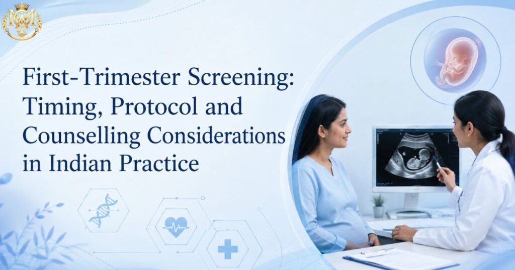
Doctor explaining first-trimester screening protocols and NT scan results to a patient
Learning blogs

First-Trimester Screening: Timing, Protocol and Counselling Considerations in Indian Practice

Quick Answer First-trimester screening for chromosomal abnormalities is optimally performed between 11+0 and 13+6 weeks of gestation. The combined screening test — NT measurement plus maternal serum free β-hCG and PAPP-A — achieves detection rates of approximately 85–90% for Down syndrome (Trisomy 21) at a 5% false-positive rate when performed within this window. NIPT (Non-Invasive Prenatal Testing) can be offered from 10 weeks onwards and carries a higher sensitivity, but does not replace diagnostic confirmation in screen-positive cases. This article covers the timing rationale, measurement standards, counselling framework, and the specific considerations that apply in Indian practice. First-trimester screening is one of the most clinically consequential steps in antenatal care — and one of the areas where clinical practice among OBGYN doctors varies most. Timing errors, suboptimal NT measurement technique, incomplete counselling, and inconsistent integration of combined screening markers are all common in routine practice, and all affect the clinical utility of the screening result. This article is a protocol-level reference. It covers the timing window and why it matters, the components of combined screening, NT measurement standards, the role of NIPT, second-line biochemical markers, and the counselling framework a practising clinician needs when delivering a screen-positive result. It also addresses the specific legal and procedural context that applies to prenatal diagnosis in India. Doctors who want structured training in prenatal screening interpretation and fetal ultrasound can explore MGA’s Fellowship in Fetal Medicine, which covers this content in depth across dedicated modules. 11–14 wk Optimal combined screening window 85–90% T21 detection rate — combined screening >99% NIPT sensitivity for T21 What this article covers Why the 11–14 week window matters — the biological rationale NT measurement — standards and common errors Combined first-trimester screening — components and interpretation NIPT — role, timing, and limitations in Indian practice Second-trimester integration — when to offer the quadruple test Screen-positive results — the counselling framework PCPNDT context — what practitioners need to know Frequently asked questions Why the 11–14 week window matters — the biological rationale The 11+0 to 13+6 week window is not arbitrary. Nuchal translucency — the fluid-filled space at the back of the fetal neck, measured on ultrasound — reaches its maximum diagnostic utility within this window. NT is physiologically present in all fetuses to a small degree, and naturally increases after 14 weeks in unaffected pregnancies as lymphatic drainage establishes. Outside this window, NT measurements are either technically not yet feasible (before 11 weeks, the fetus is too small for reliable measurement) or diagnostically unreliable (after 14 weeks, NT has already begun to resolve in normal fetuses, narrowing the gap between affected and unaffected). The CRL (crown-rump length) range within this gestational window is 45–84 mm. The CRL measurement determines whether the fetus is within the NT measurement window, not gestational age alone — this distinction matters when dates are uncertain. Practical point: When a patient presents at or near 14 weeks, do not delay referral to a better-equipped centre if your local equipment or training is not adequate for NT measurement. A NT measurement at 13+5 is still within protocol; a measurement at 14+1 is not. Missing the window means the combined test cannot be performed and the patient defaults to second-trimester screening alone, which has lower sensitivity. NT measurement — standards and common errors NT measurement is the most skill-dependent component of first-trimester screening. Poorly performed NT measurements produce unreliable risk calculations regardless of how good the serum marker results are. Measurement standards (FMF protocol) The Fetal Medicine Foundation (FMF) protocol is the widely used international standard. Key requirements: Fetal position: Sagittal section of the fetus in a neutral position — neither hyperflexed nor hyperextended. Both hyperflexion and hyperextension artificially alter the NT measurement. Image magnification: The fetal head and upper thorax should occupy the full screen. The fetus should fill at least 75% of the screen. Calipers: Placed on the inner borders of the nuchal space — not on the skin lines themselves. The horizontal part of the caliper should be barely visible against the echogenic line. Skin line vs amnion: The amniotic membrane must not be confused with the skin line. Wait for the fetus to move away from the amnion if they appear fused. Multiple measurements: Take at least three measurements and use the largest for risk calculation. Common measurement errors Error Effect on Measurement Correction Neck hyperflexion Falsely increases NT by up to 0.6 mm Reposition, wait for neutral posture Measuring on amnion, not skin Falsely increases NT Wait for fetal movement to separate amnion Calipers on outer borders Overestimates NT Use inner-to-inner caliper placement Insufficient magnification Imprecise caliper placement Head and thorax to fill >75% of screen Single measurement accepted Misses higher reproducible value Minimum 3 measurements; use the largest Combined first-trimester screening — components and interpretation Combined screening integrates three inputs into a single risk calculation for chromosomal abnormalities, primarily Trisomy 21 (Down syndrome), Trisomy 18 (Edwards syndrome), and Trisomy 13 (Patau syndrome). Three components 1. Maternal age-related background risk: The prior probability of chromosomal abnormality based on the mother’s age at the expected date of delivery — the starting point before any test modifies it. 2. Nuchal translucency (NT) measurement: Expressed as a multiple of the median (MoM) for that gestational age and CRL. An NT at or above the 95th centile (approximately ≥2.5 mm at 11 weeks, ≥3.5 mm at 13 weeks) raises the posterior risk substantially, though the precise risk change depends on the full calculation. 3. Maternal serum markers — free β-hCG and PAPP-A: Free β-hCG is elevated in Down syndrome pregnancies at 11–13 weeks (approximately 2.0 MoM). PAPP-A is reduced in Down syndrome pregnancies at 11–13 weeks (approximately 0.4–0.5 MoM). These two markers change in opposite directions in Trisomy 21, which is what gives the combined test its discriminatory power. Risk threshold and result interpretation The standard combined screening risk threshold for classifying a result as screen-positive is 1 in 300 (or 1:300) for Trisomy 21. Results above this threshold (e.g., 1:50, 1:120) are screen-positive;

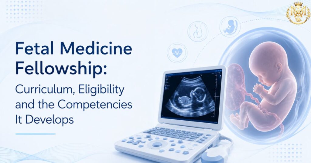
Fetal Medicine Fellowship curriculum and eligibility guide next to an ultrasound machine
Learning blogs

Fetal Medicine Fellowship: Curriculum, Eligibility and the Competencies It Develops

Quick Answer MGA’s Fellowship in Fetal Medicine is a 12-month, CPD-accredited online program covering 17 curriculum modules across fetal ultrasound, prenatal screening, fetal echocardiography, invasive procedures, high-risk pregnancy management, fetal anomaly detection, and prenatal genomics. It is designed for doctors with an obstetrics and gynaecology background — DGO, MD/MS OBG, and DNB OBGYN — who want structured subspecialty training in fetal medicine and prenatal diagnosis without pausing clinical practice. This article covers what the program specifically teaches, who is eligible, what procedural competencies it develops, and how it differs from general OBG training. If you are already practising obstetrics and considering a structured subspecialty commitment in fetal medicine, you have probably already read the persuasion-framed articles — the ones that explain what fetal medicine is and why it matters. This article serves a different purpose. It covers the specification: what MGA’s Fellowship in Fetal Medicine actually teaches module by module, who is eligible to apply, what procedural competencies it develops, and how to evaluate whether this program fits your current clinical context and career direction. For the case for why fetal medicine subspecialisation makes clinical sense for practising OBGYN doctors, MGA’s existing article on why to consider a fellowship in fetal medicine covers that well. This article picks up where that one leaves off. 17 modules Full curriculum breadth 12 months Duration — fully online CPD Accredited — international recognition What this article covers Program format and delivery — how it works for working doctors Eligibility — who the program is designed for 17-module curriculum — what is taught and in what sequence Core procedural competencies developed What this fellowship does not cover — honest scope setting How the fellowship positions you in subspecialty practice Frequently asked questions Program format and delivery — how it works for working doctors The fellowship runs for 12 months and is delivered entirely online — live sessions scheduled after OPD hours, with 100% recorded lectures available on the LMS portal for any session you cannot attend live. This format is intentional: it is built for doctors who are actively practising and cannot pause clinical work for a full-time program. The program includes weekly case clinic discussions, dedicated Q&A sessions, free study materials and an e-library with prenatal imaging guides and case references, CPD-accredited certification on completion, and one year of structured expert mentorship after the program ends. The mentorship period is not a nominal credential — it is designed to support your transition into applied fetal medicine practice once the academic year is complete. Accreditation: The fellowship is accredited by the CPD & Training Council with international continuing medical education recognition. It does not carry a university degree credential — it is a CPD-accredited professional fellowship. For doctors who specifically need a university-awarded qualification in obstetric imaging, MGA’s PG Diploma in Ultrasonography or Certificate in Advanced Ultrasound in Obstetrics and Gynecology are separate, differently-credentialled options. Eligibility — who the program is designed for The fellowship is open to eligible medical graduates. Based on the program’s clinical orientation — prenatal diagnosis, fetal ultrasound interpretation, invasive procedures, and high-risk pregnancy management — it is most directly relevant to doctors with an obstetrics and gynaecology background. Qualification Clinical Relevance to This Fellowship DGO Direct application — DGO doctors managing antenatal OPDs benefit most from structured fetal ultrasound, anomaly screening, and prenatal counselling modules MD / MS Obstetrics & Gynaecology Strong fit — builds subspecialty depth in fetal imaging, invasive procedures, and high-risk pregnancy beyond the MD/MS curriculum DNB OBGYN Strong fit — same clinical scope as MD/MS OBG; fellowship adds focused fetal medicine competency MBBS with OBG caseload Applicable — particularly for MBBS doctors working in antenatal care or managing obstetric patients in primary or secondary care settings General Physicians managing antenatal patients Relevant for the prenatal screening, high-risk pregnancy, and referral threshold modules — less directly applicable to the procedural content Check the fellowship course page for current eligibility requirements before applying — criteria may be updated periodically. 17-module curriculum — what is taught and in what sequence The 17 modules are sequenced to build from foundational knowledge through clinical application and into advanced and emerging practice areas. The outline below summarises each module’s scope. Module 1: Introduction to Fetal Medicine and Maternal-Fetal Physiology The foundational module establishes the clinical role of fetal medicine in contemporary obstetrics, covering maternal-fetal physiology, placental development and function, fetal growth, embryology, and an introduction to prenatal screening and genetic counselling frameworks. Module 2: Ultrasound and Imaging Techniques in Fetal Medicine Practical fetal ultrasound — guidelines for obstetric and gynaecological imaging, fetal biometry and growth assessment, Doppler ultrasound for fetal wellbeing, 3D/4D imaging of fetal abnormalities, amniotic fluid index assessment, placental and umbilical cord evaluation, and the application of ultrasound in chromosomal abnormality screening. Module 3: Prenatal Screening and Diagnosis First- and second-trimester aneuploidy screening protocols, NIPT clinical application and interpretation, combined screening markers, ultrasound indicators for Down syndrome and Trisomy 18, invasive diagnostic procedures (amniocentesis, CVS, fetal blood sampling), and genetic counselling communication with patients. Module 4: High-Risk Pregnancy and Maternal-Fetal Medicine Management of preeclampsia and gestational diabetes, hypertensive disorders of pregnancy including HELLP syndrome, fetal growth restriction and IUGR, multiple pregnancy diagnosis and management, premature labour prevention, antenatal corticosteroid therapy, and evidence-based monitoring of high-risk pregnancies. Module 5: Fetal Anomalies and Intervention Common fetal malformations — neural tube defects, congenital heart disease, gastrointestinal and renal defects — alongside fetal intervention indications, techniques, and outcomes, intrauterine fetal transfusion, post-fetal surgery neonatal care, and the ethical dimensions of fetal anomaly management. Module 6: Maternal-Fetal Medicine Procedures and Interventions Cordocentesis and fetal blood sampling, intrauterine treatments including laser therapy, fetal blood transfusion, and shunts, amniocentesis and CVS technique, indication, and risk counselling, and fetal wellbeing monitoring through biophysical profiles. Module 7: Ethical and Legal Aspects of Fetal Medicine Ethical dilemmas in fetal medicine practice, fetal rights versus maternal rights, decision-making frameworks for complex fetal conditions, informed consent for genetic counselling and invasive procedures, and the legal context of prenatal diagnosis in

Doctor carefully reviewing an ECG to avoid common interpretation mistakes in primary care
Learning blogs

Commonly Missed ECG Findings in Primary Care and How to Systematise Your Reading

Quick Answer Most missed ECG findings in primary care aren’t missed because the doctor doesn’t know the pattern — they’re missed because time pressure replaces a systematic read with a quick glance for the obvious. The fix is a fixed reading sequence (rate, rhythm, axis, intervals, morphology, then clinical correlation) applied to every ECG, every time — plus specific familiarity with the patterns that look deceptively unremarkable: posterior MI, de Winter T waves, Wellens syndrome, early hyperkalemia, and subtle STEMI in the presence of LBBB. This article covers both the method and the specific patterns. An ECG rarely gets misread because the underlying pattern is obscure. It gets misread because a busy OPD or emergency shift compresses a two-minute systematic read into a five-second glance for the obvious — an ST elevation that jumps out, a rate that’s clearly too fast or too slow. The findings that get missed are, almost without exception, the ones that don’t announce themselves. This article covers two things together, because they solve the same problem from different angles: a systematic reading method that structurally reduces the chance of missing something, and a specific list of the patterns most often missed even by doctors reading carefully — because they look subtle, atypical, or deceptively benign at first glance. Doctors who want to build this into a formal, structured skill can explore MGA’s Certificate in ECG Interpretation, which covers systematic reading and pattern recognition in depth. 7 steps In a systematic reading method 8 patterns Most frequently missed in primary care 0 shortcuts The method works because it skips none What this article covers Why ECG misses happen even when doctors know the patterns A systematic reading method you can apply to every ECG Eight commonly missed findings — and why each one hides How to build reading speed without cutting corners Frequently asked questions Why ECG misses happen even when doctors know the patterns Ask most doctors whether they know what a de Winter T wave pattern looks like, or what Wellens syndrome represents, and many will say yes. Knowledge isn’t usually the gap. What breaks down in practice is application under pressure — a busy OPD queue, an emergency department at capacity, or simple pattern fatigue after reading dozens of unremarkable ECGs in a row. Three specific failure modes account for most misses in primary care and general emergency settings: Single-glance triage: Scanning for the one obvious abnormality — a clear ST elevation or an obviously irregular rhythm — and stopping there, rather than completing a full systematic read regardless of what’s found early. Anchoring on a normal-looking baseline: Several high-stakes patterns (posterior MI, Wellens syndrome, early hyperkalemia) present with a standard 12-lead ECG that looks largely unremarkable at first pass, because the abnormality is subtle, reciprocal, or in a lead group that’s easy to underweight. Isolated reading without clinical correlation: Reading the ECG as a standalone image rather than integrating it with the presenting complaint. A patient who was pain-free at the time of the ECG but describes classic exertional chest pain in the history needs the ECG read with that context in mind — a resting, asymptomatic ECG can look deceptively reassuring in exactly this scenario. A systematic reading method you can apply to every ECG The single highest-value habit in ECG interpretation is applying the same fixed sequence to every ECG, regardless of how obvious or unremarkable it appears at first glance. The sequence below is a standard, widely taught structure — the value isn’t in any single step being novel, it’s in never skipping one. 1. Rate Establish the ventricular rate first — both to flag obvious tachycardia or bradycardia and because rate context changes how you interpret everything that follows. 2. Rhythm Determine whether the rhythm is regular or irregular, and whether it’s sinus. This step alone catches atrial fibrillation, flutter, and most significant arrhythmias — but only if performed deliberately rather than assumed from a “normal-looking” rate. 3. Axis Assess the QRS axis. Axis deviation can point toward chamber hypertrophy, conduction abnormalities, or prior infarction — findings that are easy to skip because axis assessment feels like an extra step rather than an obviously urgent one. 4. Intervals Measure the PR interval (looking for heart block), QRS duration (looking for bundle branch block or conduction delay), and QT interval (looking for prolongation relevant to arrhythmia risk and drug interactions). Each interval answers a different clinical question — none should be skipped because another looks normal. 5. P wave and QRS morphology Examine P wave shape and consistency (atrial enlargement, ectopic atrial rhythms) and QRS morphology (bundle branch blocks, ventricular hypertrophy, pathological Q waves indicating prior infarction). 6. ST segment and T wave This is where most acute ischemic findings live — and where most subtle misses happen. Assess every lead group systematically for ST elevation, ST depression, and T wave changes, including reciprocal changes in leads that aren’t the “obvious” ones for the clinical presentation. 7. Clinical correlation Integrate the ECG findings with the presenting complaint, risk factors, and timing of symptoms. An ECG is a snapshot — a normal or near-normal ECG in a patient with a highly suggestive history does not rule out a significant cardiac event, particularly if the ECG was taken between symptomatic episodes. Build a formal, structured ECG interpretation skill MGA’s Certificate in ECG Interpretation — systematic reading, rhythm recognition, ischemia patterns View Program Eight commonly missed findings — and why each one hides Finding What It Looks Like Why It’s Missed Posterior MI ST depression and tall R waves in V1–V3 — a reciprocal, mirror-image pattern rather than direct ST elevation No ST elevation is visible on the standard 12-lead; the abnormality is a mirror pattern in anterior leads, easily read as “unremarkable” De Winter T wave pattern Upsloping ST depression with tall, symmetric T waves in the precordial leads Absence of ST elevation despite representing an acute proximal LAD occlusion equivalent — easily read as non-specific change Wellens syndrome Deeply

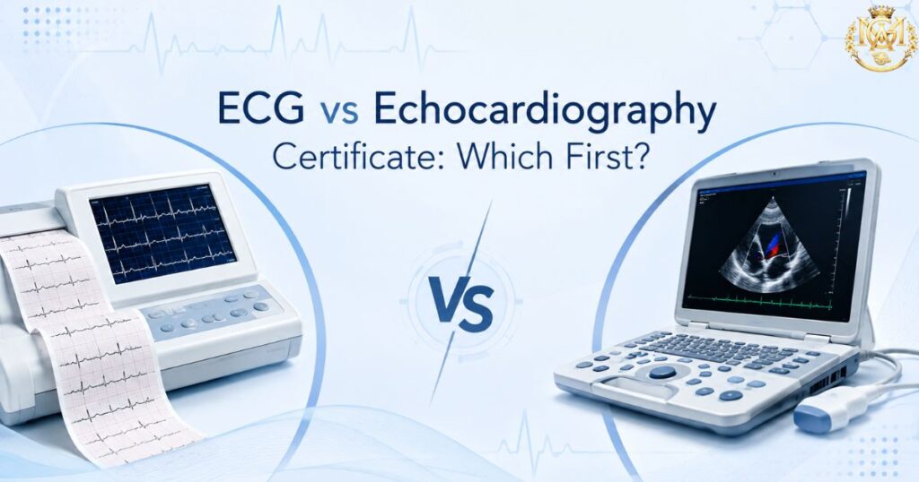
Doctor comparing clinical skills for a certificate in ECG interpretation and echocardiography
Learning blogs

Certificate in ECG Interpretation vs Certificate in Echocardiography: Which Cardiac Skill First?

Quick Answer For most MBBS doctors, general physicians, and emergency practitioners, ECG interpretation is the stronger first choice — it requires no equipment beyond what most clinics already have, applies to nearly every patient encounter involving chest pain or palpitations, and builds a diagnostic reflex you’ll use daily. Echocardiography is the better first choice specifically if you already work alongside an ultrasound machine or want to add bedside cardiac imaging to your practice. Many doctors eventually complete both — the sequencing question is really about which skill pays off faster in your specific setting. Cardiac skill-building is rarely an all-or-nothing decision. Most doctors don’t have the time or need for a full cardiology fellowship, but they do encounter chest pain, palpitations, and breathlessness often enough that a focused cardiac skill genuinely changes how confidently they manage a patient. The real question most doctors face isn’t “should I learn cardiology” — it’s “which specific skill should I build first.” MGA’s Certificate in ECG Interpretation and Certificate in Echocardiography are two distinct answers to that question. This article compares them directly — what each actually teaches, what equipment and setting each assumes, and which one to prioritise based on your current practice. If you’re still exploring the full range of cardiology course formats available after MBBS, our cardiology courses after MBBS guide covers that broader landscape; this article is specifically about sequencing these two certificates. 2 skills Compared for sequencing, not ranked Zero vs Equipment Core practical difference Daily use Both apply to routine OPD and ER work What this guide covers What the Certificate in ECG Interpretation covers What the Certificate in Echocardiography covers Direct comparison: equipment, learning curve, and daily use Who should learn ECG interpretation first Who should learn echocardiography first Can you build both skills — and in what order? Frequently asked questions What the Certificate in ECG Interpretation covers The Certificate in ECG Interpretation is built around a single, focused competency: reading a 12-lead ECG accurately and confidently, in the time pressure of a real clinical encounter — not in the unhurried setting of a textbook. Core content areas ECG fundamentals and lead placement: Cardiac electrical conduction, the relationship between the 12 leads and cardiac anatomy, and correct electrode placement — the foundation that makes every subsequent reading accurate rather than technically flawed from the start. Systematic reading method: A structured, repeatable approach to reading any ECG — rate, rhythm, axis, intervals, and morphology in a fixed sequence — designed to replace ad hoc pattern-spotting with a method that catches findings a rushed read misses. Rhythm recognition: Identifying normal sinus rhythm and the common arrhythmias encountered in general and emergency practice — atrial fibrillation, atrial flutter, supraventricular tachycardia, ventricular ectopy, and heart blocks. Ischemia and infarction patterns: Recognising ST elevation, ST depression, T wave inversion, and Q wave changes, along with the less obvious patterns that carry high clinical stakes — findings that are easy to miss under time pressure but change immediate management when caught. Clinical correlation and escalation thresholds: When an ECG finding warrants immediate referral, when it can be managed and monitored, and when it’s a benign variant — the judgment layer that separates confident interpretation from technically correct but clinically unhelpful reading. What the daily use looks like ECG interpretation requires no equipment beyond a standard ECG machine — already present in most clinics, OPDs, and emergency departments. The skill applies immediately to any patient presenting with chest pain, palpitations, breathlessness, or syncope, making it one of the most frequently used cardiac skills in general practice. View the Certificate in ECG Interpretation Focused, applied cardiac skill training · Eligible doctors View Program What the Certificate in Echocardiography covers The Certificate in Echocardiography is built around bedside cardiac ultrasound — a fundamentally different skill from ECG in that it involves image acquisition and spatial interpretation, not just waveform reading. Core content areas 2D echo fundamentals and transducer technique: Basic ultrasound physics as applied to cardiac imaging, transducer positioning, and the standard transthoracic echocardiography (TTE) windows — parasternal, apical, subcostal, and suprasternal views. Chamber assessment: Systematic evaluation of the four cardiac chambers — size, wall thickness, and gross structural assessment — building the visual vocabulary needed to recognise normal versus abnormal anatomy. Basic valve assessment: Visual assessment of valve structure and motion, and recognising overt valvular abnormalities — a foundational level of valve evaluation rather than comprehensive Doppler-based quantification. LV function and ejection fraction estimation: Visual (“eyeballing”) assessment of left ventricular contractility and a basic approach to estimating ejection fraction — one of the most clinically useful bedside echo skills, directly relevant to heart failure and acute cardiac presentations. When to refer for formal echocardiography: Recognising the boundary between what a focused certificate-level skill can answer and when a comprehensive echocardiographic study from a cardiologist or trained sonographer is required. What the daily use looks like Echocardiography requires access to an ultrasound machine — a real practical constraint that ECG interpretation does not carry. The skill is most immediately useful in settings where cardiac imaging equipment is already available: hospitals, diagnostic centres, and clinics with point-of-care ultrasound capability. View the Certificate in Echocardiography Bedside cardiac imaging skill training · Eligible doctors View Program Direct comparison: equipment, learning curve, and daily use Factor ECG Interpretation Echocardiography Equipment required Standard ECG machine — near-universal availability Ultrasound machine — not present in every clinic Skill type Waveform reading and pattern recognition Image acquisition + spatial interpretation Learning curve Faster — no hand-eye probe coordination needed Longer — image quality is skill-dependent Frequency of clinical use Very high — any chest pain/palpitation presentation High where equipment exists, low otherwise Standalone diagnostic value High — often decisive on its own for acute presentations High but frequently used alongside other findings Typical settings OPD, emergency department, rural/primary care Hospitals, diagnostic centres, imaging-equipped clinics Best first skill for General physicians, emergency doctors, rural practitioners Doctors already working alongside ultrasound equipment Course duration, fees, and specific eligibility should be verified on each

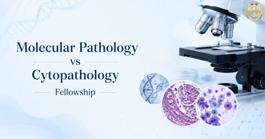
Pathologists comparing subspecialties to choose between a fellowship in molecular pathology and cytopathology
Learning blogs

Fellowship in Molecular Pathology vs Cytopathology: Choosing a Diagnostic Subspecialty

Quick Answer Molecular pathology and cytopathology are both diagnostic subspecialties within pathology, but they involve fundamentally different daily work. Molecular pathology centres on nucleic acid-based testing — PCR, sequencing, biomarker analysis — that informs targeted therapy and precise disease classification, often performed at the bench with limited direct patient contact. Cytopathology centres on interpreting cellular samples — FNAC, exfoliative cytology, effusion fluids — often with same-day, rapid-turnaround reporting and, in many practices, direct involvement in performing the aspiration itself. Choose based on which kind of diagnostic work suits your interest and the kind of laboratory or hospital setting you want to build a career in — not on which sounds more advanced. Every MD Pathology or DNB Pathology holder eventually faces a subspecialty fork. General diagnostic pathology gives broad competence, but career depth and consultant-level differentiation increasingly come from a defined subspecialty focus. Two of the most commercially and clinically relevant options are molecular pathology and cytopathology — and they are frequently confused as adjacent when they are, in practice, quite different disciplines. This article compares MGA’s Fellowship in Molecular Pathology and Fellowship in Cytopathology directly — what each subspecialty actually involves day to day, what skill set each builds, and which one is the better fit depending on your existing practice and career direction. If you are still evaluating pathology as a specialty more broadly, our Fellowship in Pathology overview is the right starting point before narrowing to a subspecialty. 2 paths Distinct diagnostic subspecialties compared Bench vs bedside Core practice difference 1 year Typical fellowship duration What this guide covers What the Fellowship in Molecular Pathology covers What the Fellowship in Cytopathology covers Direct comparison: daily practice, skills, and career settings Who should choose molecular pathology Who should choose cytopathology Can you build competency in both? Frequently asked questions What the Fellowship in Molecular Pathology covers Molecular pathology is the diagnostic discipline concerned with detecting disease at the level of nucleic acids and molecular biomarkers rather than tissue architecture or cell morphology alone. It has become central to modern oncology, infectious disease diagnostics, and inherited disease testing because treatment decisions increasingly depend on molecular characterisation rather than histological appearance alone. Core content areas Molecular techniques and principles: Polymerase chain reaction (PCR) and its variants, nucleic acid extraction, Sanger sequencing, and the principles of next-generation sequencing (NGS) form the technical foundation. Understanding how these techniques work — not just what they report — is what allows a pathologist to interpret results critically rather than simply transcribe them. Molecular oncology: Biomarker testing that guides targeted cancer therapy — EGFR, ALK, KRAS, BRCA, and similar actionable mutations — along with the tumour classification systems that increasingly integrate molecular findings alongside histology. This is the area of molecular pathology with the most direct clinical impact on treatment selection. Molecular infectious disease diagnostics: PCR-based pathogen detection, viral load quantification, and resistance testing — applications that have expanded substantially in Indian laboratories following increased demand for rapid, sensitive infectious disease diagnostics. Inherited disease and genetic testing principles: The basis of testing for inherited conditions, carrier screening principles, and the interpretation framework for genetic variants — foundational knowledge relevant to laboratories offering genetic testing services. Quality assurance and test validation: Molecular testing carries specific quality assurance requirements — assay validation, proficiency testing, and result reporting standards — that differ substantially from morphology-based diagnostic work. This is a curriculum area with direct laboratory management relevance. What the daily work looks like Molecular pathology practice is largely laboratory- and bench-based. The pathologist’s role centres on test selection, result interpretation in clinical context, and quality oversight of the molecular testing process — with limited direct patient contact compared to procedural diagnostic disciplines. It suits doctors who are drawn to the technical and interpretive precision of nucleic acid-based diagnostics. View the Fellowship in Molecular Pathology CPD accredited · Evening classes after OPD · Eligible doctors View Program What the Fellowship in Cytopathology covers Cytopathology is the diagnostic discipline concerned with interpreting individual cells and small cell clusters obtained from fluid samples, scrapings, or fine needle aspiration — rather than the tissue architecture examined in histopathology. It is one of the most immediately clinically applied pathology subspecialties, frequently involving rapid, same-visit diagnostic decisions. Core content areas Fine needle aspiration cytology (FNAC): Technique and interpretation for aspirating and evaluating superficial masses — thyroid nodules, lymph nodes, breast lumps, and salivary gland masses are the most common. FNAC training covers both the aspiration technique itself and the cytological interpretation of the resulting sample. Gynecological cytology: Cervical cytology screening and interpretation, including the recognition of pre-malignant and malignant cervical changes — a high-volume diagnostic area with direct population health relevance in cervical cancer screening programmes. Non-gynecological exfoliative cytology: Interpretation of cells shed into body fluids — sputum, urine, pleural and peritoneal effusions, and cerebrospinal fluid. This is a substantial component of general cytopathology practice, particularly in the evaluation of effusions for malignancy. Rapid on-site evaluation principles: The practice of assessing specimen adequacy at the time of collection — a skill that directly reduces the need for repeat procedures and speeds diagnostic turnaround, particularly relevant when aspirations are image-guided. Ancillary technique correlation: Understanding how immunocytochemistry and, where relevant, molecular testing on cytology specimens integrate with cytomorphological interpretation to reach a definitive diagnosis. What the daily work looks like Cytopathology combines interpretive laboratory work with, depending on practice setting, direct involvement in specimen collection through FNAC. Reporting turnaround is frequently rapid — same-day or next-day in many settings — and the diagnostic reasoning is built around recognising cellular patterns rather than tissue architecture. It suits doctors who want a subspecialty with both a procedural and interpretive dimension. View the Fellowship in Cytopathology CPD accredited · Evening classes after OPD · Eligible doctors View Program Direct comparison: molecular pathology vs cytopathology Factor Molecular Pathology Cytopathology Core diagnostic basis Nucleic acids, biomarkers, molecular alterations Cellular morphology in fluid and aspirate samples Patient contact Minimal — primarily bench and interpretive work Variable — FNAC often involves direct

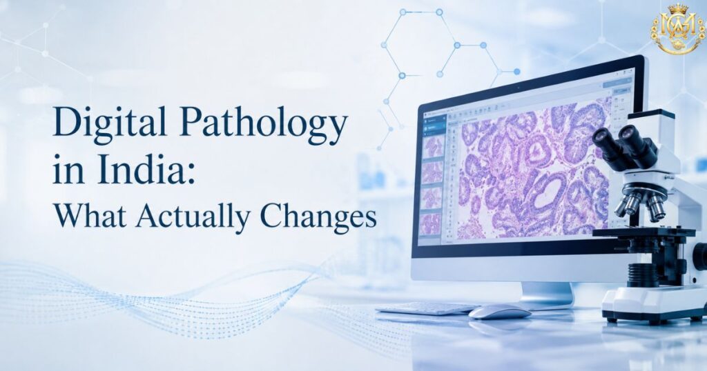
Pathologist reviewing a whole slide image on a screen representing digital pathology in India
Learning blogs

Digital Pathology and AI-Assisted Diagnostics: What Changes in the Indian Laboratory

Quick Answer Digital pathology replaces the glass slide and microscope with a scanned digital image viewed and reported on a screen — enabling remote sign-out, easier consultation, and integration with AI-assisted image analysis tools. In Indian laboratories, the practical changes are showing up in three areas: reporting workflow (screen-based review, digital annotation, remote second opinions), quality assurance (new validation requirements before clinical deployment), and turnaround time (faster in some workflows, unchanged or slower in others during the transition period). AI tools currently assist with specific, narrow tasks — quantification, screening triage, pattern flagging — rather than replacing pathologist judgment on final diagnosis. Digital pathology has been discussed as an imminent transformation for over a decade. What has actually changed in Indian laboratory practice is more incremental and more specific than the broader “AI will transform pathology” framing suggests. This article sets aside the hype and looks at what digital pathology and AI-assisted diagnostics concretely alter for a working pathologist — in reporting workflow, quality assurance, and turnaround time — and what remains unchanged. This is a technology-and-practice piece, not a career guide. For pathology subspecialty and career pathway content, see our comparison of Fellowship in Molecular Pathology vs Cytopathology, and MGA’s Fellowship in Pathology for the foundational program. Growing India’s digital pathology adoption, per market analysts Scanners Largest segment of India’s digital pathology market Narrow AI Current role: quantification & triage, not diagnosis Market growth figures for India’s digital pathology sector vary substantially across research providers depending on methodology and market definition; directional growth is consistently reported, though we avoid citing a single disputed figure here. What this article covers What digital pathology actually is — and is not What changes in reporting workflow Where AI-assisted diagnostics genuinely help today Where AI does not replace pathologist judgment Telepathology and remote reporting in the Indian context Quality assurance and validation implications Turnaround time: what actually speeds up, what doesn’t What this means for pathologist skill requirements Frequently asked questions What digital pathology actually is — and is not Digital pathology, at its core, is the digitisation of the glass slide. A whole slide imaging (WSI) scanner captures a high-resolution digital image of the entire slide, which the pathologist then views, annotates, and reports on a computer screen rather than through a microscope eyepiece. The digital image can be stored, transmitted, shared for consultation, and — where validated systems are in place — analysed by computational tools. It is worth being precise about what this is not. Digital pathology is not, by itself, a diagnostic method — it is an image acquisition and viewing method. The diagnostic reasoning remains the pathologist’s, whether performed on glass or on screen. AI-assisted diagnostics is a separate, additional layer that can be built on top of digitised images — but digitisation and AI analysis are not the same thing, and a laboratory can digitise its workflow without deploying any AI tools at all. In India, scanner devices currently represent the largest single segment of digital pathology market activity, reflecting where laboratories are actually investing — in the acquisition and viewing infrastructure — ahead of widespread AI deployment. Institutions such as KIMS Odisha have publicly commissioned whole slide scanning systems as part of routine cancer diagnosis workflows, illustrating the practical, incremental nature of adoption rather than a wholesale technology replacement. What changes in reporting workflow Screen-based sign-out replaces the microscope for primary review The most direct workflow change is that primary diagnostic review shifts from eyepiece to screen. This changes ergonomics, requires monitor calibration standards to preserve colour and detail fidelity, and requires a different visual scanning discipline — moving across a digital slide is not identical to moving a physical slide under a microscope, and pathologists transitioning to digital sign-out typically undergo a validation period comparing their digital and glass-slide diagnostic concordance before full digital transition. Digital annotation and case marking Digital slides allow annotation tools — marking regions of interest, measuring dimensions directly on the image, and flagging areas for a colleague’s review — that are more precise and shareable than physical slide marking. This is a genuine workflow improvement for teaching, multidisciplinary case review, and documentation. Remote consultation and second opinions Perhaps the most immediately valuable change: a digitised slide can be shared instantly with a specialist anywhere, without physically shipping the glass slide. For a complex or unusual case requiring subspecialty input, this collapses what was previously a multi-day courier process into a same-day digital consultation. This capability underpins much of the telepathology discussion in the Indian context (covered below). Integration with laboratory information systems Digital pathology platforms increasingly integrate with laboratory information systems (LIS), linking the digital image directly to the patient record, prior reports, and relevant clinical data — reducing the manual cross-referencing that glass-slide workflows require. Where AI-assisted diagnostics genuinely help today The realistic, current-state role of AI in pathology is narrow and task-specific — not the general diagnostic replacement that popular framing sometimes suggests. The applications with genuine, demonstrated value fall into a few defined categories. Quantification tasks Counting mitotic figures, quantifying Ki-67 proliferation index, and measuring immunohistochemistry staining intensity are tasks that are time-consuming and subject to inter-observer variability when performed manually. AI-assisted quantification tools perform these specific counting and measurement tasks with good reproducibility, and are among the most mature and clinically adopted AI applications in pathology internationally. Screening and triage in high-volume cytology In cervical cytology screening, AI-assisted pre-screening can flag slides most likely to contain abnormal cells for prioritised pathologist review — a triage function that helps manage high case volumes rather than a diagnostic replacement. The pathologist still makes the final diagnostic call on flagged and unflagged material according to laboratory protocol. Pattern detection as a second check AI tools trained to flag specific patterns — certain tumour morphologies, particular infectious organisms, specific staining patterns — can serve as a second-check overlay, similar in concept to spell-check: a prompt for the pathologist to look again at a specific region, not a replacement diagnosis.

Scroll to Top