Medical Global Academy

Learning blogs

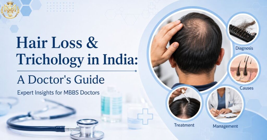
Doctor examining a patient's scalp representing a clinical guide to hair loss and trichology in India
Learning blogs

Hair Loss & Alopecia in India: When to Treat, When to Refer, and the Rise of Trichology

📅 Updated September 2026 ⏰ 9 min read 🩺 MBBS & General Physicians 🌎 Dermatology Cluster — Day 7 ⚡ Quick Answer What’s the difference between trichology and dermatology for hair loss? Dermatology is the established medical specialty that already covers hair and scalp disorders as part of comprehensive skin, hair, and nail care — most alopecia in India is diagnosed and managed by dermatologists. Trichology is a narrower, emerging focus area on hair and scalp specifically, without a single standardised medical degree behind it in India the way MD Dermatology has. For most patients and most doctors, dermatology remains the credentialed route; trichology is best understood as a specialised interest area within it, not a separate, equivalent qualification. Hair loss consultations are becoming a routine part of general practice and dermatology OPDs across India, and “trichology” is a term doctors are hearing more often — in clinic branding, in continuing education circles, and increasingly in patient questions. But what trichology actually is, how it differs from dermatology, and whether it’s a credible career direction for a doctor are questions that don’t get answered clearly very often. This guide covers the common types of alopecia you’ll see in practice, what trichology genuinely is (and isn’t), general management principles before referral, the systemic conditions hair loss can signal, and an honest look at trichology as a career direction. Why Hair Loss Consultations Are Rising in India A few converging factors are driving more patients to seek treatment for hair loss rather than accept it as inevitable. Awareness has grown substantially — patients now recognise androgenetic alopecia, telogen effluvium, and alopecia areata as distinct, sometimes treatable conditions, rather than lumping all hair loss together as “just getting older.” Younger patients are presenting earlier too, often prompted by visible early thinning that previous generations may have simply lived with. Urban lifestyle factors — chronic stress, irregular sleep, nutritional patterns, and pollution exposure — are frequently raised by patients themselves as suspected contributors, even when the actual cause is more specifically hormonal or genetic. Alongside this, cosmetic and hair-restoration clinics have expanded significantly in tier-1 and tier-2 cities, increasing both patient awareness and the range of places patients might first seek help — not always from a qualified dermatologist. Common Types of Alopecia: A Quick Differentiator Three patterns account for the large majority of hair loss presentations in general practice and dermatology OPDs. Androgenetic Alopecia Gradual, patterned hair thinning — temporal recession and vertex thinning in men, diffuse crown thinning in women. Genetically and hormonally driven (androgen-mediated), typically progressive without intervention. Genetic/hormonal Progressive Telogen Effluvium Diffuse shedding, typically emerging 2–3 months after a triggering event — illness, surgery, childbirth, severe stress, or rapid weight loss. Usually self-limiting once the underlying trigger resolves. Usually self-resolving Trigger-linked Alopecia Areata Autoimmune, well-demarcated round or oval patches of hair loss, can affect any hair-bearing area. Course is unpredictable — some cases resolve spontaneously, others progress or recur. Autoimmune Unpredictable course The distribution pattern and history usually point to the diagnosis quickly: gradual and patterned suggests androgenetic; sudden and diffuse following a stressor suggests telogen effluvium; sharply demarcated round patches suggest alopecia areata. What Is Trichology, and How Is It Different From Dermatology? Trichology, broadly, is the study of hair and scalp health and disorders. Internationally, it emerged historically as a cosmetology-adjacent field, and it has become increasingly medicalised in recent years as interest in hair restoration has grown. Dermatology, by contrast, is an established, regulated medical specialty in India — an MD Dermatology programme already includes hair and scalp disorders as one of its core domains, alongside skin and nail conditions. A qualified dermatologist is trained to diagnose and manage the full range of alopecia types, including the ones that require careful clinical judgement, like distinguishing early scarring alopecia from a more benign pattern. The practical complication in the Indian market is that “trichology” isn’t attached to a single, standardised, nationally regulated medical degree the way MD Dermatology is. Trichology-branded clinics range widely — some are run by qualified dermatologists who’ve developed a specific interest in hair disorders, others blend cosmetic services like PRP and hair transplant coordination with less rigorous diagnostic oversight, and the line between “trichologist” and “hair cosmetologist” isn’t always clear to patients. ⚠️ For general physicians: this is exactly why referring suspected alopecia areata, any hair loss with signs of scarring or active inflammation, or diagnostically unclear presentations to a qualified dermatologist matters more than referring to a “hair clinic” by name alone. Credentials matter more than branding. General Management Principles Before Referral A structured initial approach helps most patients, even before a specialist is involved. History first. Establish onset (sudden vs gradual), pattern (diffuse vs localised), family history of pattern baldness, any recent illness, surgery, severe stress, or significant dietary change, and — in women — menstrual regularity and any signs suggestive of excess androgen (irregular cycles, acne, hirsutism), which raises the possibility of an underlying condition like PCOS. Basic examination. Note the distribution pattern, look for scarring or active inflammation on the scalp, and a gentle hair-pull test can help gauge whether shedding is currently active. General first-line guidance. For telogen effluvium, reassurance is often the most valuable initial intervention — most cases resolve within months once the trigger is addressed, and patients benefit from understanding this is usually temporary. For androgenetic alopecia, setting realistic expectations about gradual, sustained management (rather than a quick fix) helps prevent frustration and reduces the temptation to try unregulated over-the-counter products. When not to delay referral. Alopecia areata, any pattern suggesting scarring alopecia, rapidly progressive diffuse loss, or diagnostic uncertainty should be referred to a dermatologist without extended trial-and-error in general practice — early specialist involvement can meaningfully affect outcomes, particularly for scarring processes where lost follicles may not recover. When Hair Loss Signals a Systemic Issue Hair loss is sometimes the presenting symptom of a broader systemic issue, which makes a basic workup worthwhile in the right clinical context. Systemic Cause Typical Clue Relevant
Medical Officer vs Junior Resident after MBBS: clinical job comparison and career path
Learning blogs

Medical Officer vs Junior Resident: Which Clinical Job First After MBBS?

Completing MBBS and internship is a major transition. For many doctors, the next question is not simply “What job can I get after MBBS?” but “Which clinical role will give me the best start to my career?” Two commonly considered options are Medical Officer (MO) and Junior Resident (JR). Both can provide hands-on patient-care experience, but they differ in purpose, structure, supervision, departmental exposure, employment conditions and long-term career relevance. The important point is that there is no universally better role. A Junior Resident position in a high-volume teaching hospital may offer structured specialty exposure, while a Medical Officer position may provide broader responsibility and immediate employment. The actual job description matters more than the designation alone. Medical Officer vs Junior Resident: What’s the Difference? What Is a Medical Officer? A Medical Officer is a licensed doctor responsible for providing clinical services within the scope defined by the employer and appointment. Medical Officers may work in: Government hospitals Private and corporate hospitals Primary and community health facilities Emergency services Clinics Public-health programmes Institutional healthcare settings Their responsibilities can include patient assessment, initial management, documentation, referrals, emergency care and coordination with other healthcare professionals. The role can therefore be quite broad. A Medical Officer working in a busy emergency department may have a very different working day from one working in a primary-care or public-health setting. What Is a Junior Resident? A Junior Resident generally works within a hospital-based clinical hierarchy and combines patient-care responsibilities with professional learning. However, the term Junior Resident is not used identically everywhere. Some institutions use it for doctors enrolled in postgraduate training, while others recruit non-PG Junior Residents on fixed-term appointments. This distinction is particularly important when evaluating a job advertisement. A non-PG JR position should not automatically be treated as equivalent to being enrolled in an MD/MS/DNB programme. Medical Officer vs Junior Resident at a Glance Factor Medical Officer Junior Resident Primary focus Clinical service Clinical service + structured learning Typical setting Hospital, clinic, health facility or programme Usually hospital/department based Specialty exposure Depends on posting Often department-specific Supervision Depends on employer Usually within a clinical hierarchy Tenure Permanent, contractual or fixed-term Often defined by appointment/notification Career value Clinical experience and employment Clinical exposure and potential preparation for specialization The designation alone does not tell you the quality of the experience. Who Is Eligible for Medical Officer and Junior Resident Jobs After MBBS? MBBS and Internship Requirements For most clinical appointments after MBBS, doctors need to have completed their medical qualification and the required internship. The compulsory rotating internship is an important transition between medical education and independent professional practice. Completion of internship is also commonly linked to the registration requirements applicable to medical practice. Medical Registration Requirements Doctors should verify the registration requirements specified in the particular recruitment notification or appointment. For an MBBS graduate, this generally means having the appropriate medical registration required to practise and meeting any registration conditions specified by the employer. Age and Experience Requirements There is no single age or experience rule covering every Medical Officer or Junior Resident position. A fresh MBBS graduate may be eligible for some entry-level positions, while other Medical Officer vacancies can ask for previous clinical experience. Similarly, Junior Resident recruitment can have institution-specific conditions regarding internship completion, previous residency and other eligibility criteria. Therefore, always read the actual recruitment notification rather than relying on a generic eligibility statement. How Eligibility Can Differ by Institution Government medical colleges, central institutions, state health departments and private hospitals can use different recruitment systems. Before applying, check: MBBS recognition requirements Internship completion date Registration requirements Age limit Previous JR experience Required experience Selection method Appointment tenure Department allocation What Does a Medical Officer Do? The Medical Officer’s exact responsibilities depend on the institution and posting. Patient Assessment and Initial Management Medical Officers commonly assess patients, take relevant clinical histories, perform examinations, identify urgent problems and initiate appropriate management within their role. In emergencies, the doctor must recognize deterioration quickly, initiate appropriate emergency measures and escalate to senior or specialist support when required. OPD and Emergency Responsibilities Depending on the posting, an MO may work in outpatient services, emergency departments or both. Typical responsibilities may include: Initial patient assessment Clinical documentation Basic investigations and interpretation Initial treatment Emergency stabilization Referral to appropriate specialists Follow-up planning The key difference is that an MO’s responsibilities are determined by the actual job description and facility, rather than by the title alone. Inpatient Clinical Responsibilities Where Medical Officers are involved in inpatient services, responsibilities may include reviewing admitted patients, monitoring clinical progress, coordinating investigations, communicating with specialists and maintaining appropriate medical records. Referrals and Coordination With Specialists A Medical Officer may often serve as an important link between the patient and specialist team. Good referral practice requires communicating the relevant clinical history, examination findings, investigations, reason for referral and urgency clearly. Documentation and Medical Records Clinical documentation is not simply administrative paperwork. Accurate and timely records support: Continuity of care Communication between clinicians Clinical decision-making Quality improvement Audit and research Medico-legal accountability For a young doctor, developing disciplined documentation habits early is an important professional skill. Public Health and Community-Based Roles Medical Officer positions can also provide exposure to community medicine and public-health programmes, particularly in government and community-based settings. This can include preventive healthcare, screening, health education, programme implementation and coordination with public-health teams. What Does a Junior Resident Do? Department-Based Patient Care Junior Residents generally work within a specific hospital department or clinical service. This can provide exposure to a defined patient population and allow the doctor to become familiar with departmental workflows, investigations, treatment protocols and procedures. Inpatient and Emergency Duties JRs may participate in: Admissions Ward rounds Patient monitoring Emergency duties Clinical documentation Investigations Treatment coordination Night duties The workload varies significantly between hospitals and departments. Ward Rounds and Case Presentations Ward rounds are an important part of hospital-based clinical learning. A resident may need to review the patient’s current condition, investigations, treatment response and outstanding

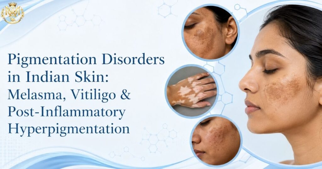
Clinical comparison of melasma, vitiligo and post-inflammatory hyperpigmentation presentations in Indian skin showing symmetrical facial patches, depigmented lesions and localized dark marks
Learning blogs

Pigmentation Disorders in Indian Skin: Melasma, Vitiligo & Post-Inflammatory Hyperpigmentation

Pigmentation Disorders in Indian Skin: Melasma, Vitiligo & Post-Inflammatory Hyperpigmentation Pigmentation Disorders in Indian Skin require a different clinical approach because pigmentation can present differently and carry a higher risk of treatment-related complications in Fitzpatrick IV–VI skin. Melasma, vitiligo, and post-inflammatory hyperpigmentation are common concerns in Indian dermatology practice, and understanding their presentation, general management principles, and referral thresholds is important for doctors treating skin of colour. This guide covers how these three conditions present specifically in Indian skin, general management principles, and when referral to a dermatologist is warranted. Why Pigmentation Disorders Present Differently in Indian Skin Skin of colour — generally defined as Fitzpatrick types IV through VI — has a higher baseline melanocyte activity than lighter skin types. This means the same inflammatory trigger that might cause temporary redness in fair skin often produces visible, sometimes persistent pigmentation change in darker skin. This has two practical consequences that shape everything else in this guide. First, pigmentation disorders in Indian patients tend to be more visually prominent and psychologically distressing, because pigmentary change is more noticeable against darker skin. Second, treatments that work well in Fitzpatrick I–III skin carry meaningfully higher risk in IV–VI skin. Laser and light-based treatments in particular carry a documented higher risk of triggering post-inflammatory hyperpigmentation when used in darker skin types, which is why treatment protocols need to be adapted rather than directly transferred from Western dermatology literature. Understanding this distinction is the starting point for managing all three conditions covered below. Melasma: Presentation, Triggers, General Management Melasma is one of the most common pigmentation complaints in Indian dermatology clinics, disproportionately affecting women, though it does occur in men as well. It presents as symmetrical, brownish-grey patches typically on the cheeks, forehead, upper lip, and chin. Common Triggers Sun exposure is the single most consistent aggravating factor, made worse by India’s high UV index for most of the year. Hormonal factors — pregnancy, oral contraceptives, hormone replacement therapy — are strongly associated triggers, which is why melasma is sometimes referred to as the “mask of pregnancy.” Genetic predisposition plays a role too; a family history of melasma is common among affected patients. General Management Principles Photoprotection is the foundation of any melasma management plan, and it isn’t optional — without consistent sun protection, other treatments have limited durability. Broad-spectrum sunscreen with both UVA and UVB coverage, reapplied through the day, is a baseline recommendation for every patient. Topical depigmenting agents are typically first-line treatment, used under proper dermatological guidance to balance efficacy against irritation risk, since irritation itself can worsen pigmentation in darker skin. Procedural treatments such as chemical peels and laser therapy are generally considered second-line, used after topical approaches have been tried, and require particular caution in Fitzpatrick IV–VI patients due to the elevated risk of triggering post-inflammatory hyperpigmentation as a side effect of the treatment itself. This is a case where the treatment for one pigmentation disorder can inadvertently cause another. Melasma is a chronic, relapsing condition. Setting realistic expectations with patients early — that the goal is improvement and control rather than permanent cure — prevents frustration and reduces the temptation to pursue aggressive procedural treatments that carry higher risk in darker skin. Vitiligo: Presentation, Patient Counselling, When to Refer Vitiligo presents as well-demarcated, depigmented (not hypopigmented) patches caused by autoimmune destruction of melanocytes. In Indian skin, the contrast between depigmented patches and surrounding normal skin is stark, which makes the condition highly visible and, in many cases, socially significant in ways that go well beyond the medical impact. Recognising Vitiligo Vitiligo typically appears first on the face, hands, feet, and areas prone to friction or trauma — a phenomenon known as the Koebner response, where new lesions develop at sites of skin injury. Patches are usually symmetrical and can be segmental (following a specific body region) or non-segmental (widespread and progressive). Unlike post-inflammatory hyperpigmentation or melasma, vitiligo patches lack all pigment rather than simply appearing lighter. Patient Counselling Vitiligo carries disproportionate social stigma in the Indian context, sometimes affecting marriage prospects, social participation, and mental health. Effective counselling addresses this reality directly rather than only the dermatological aspect. Patients benefit from understanding that vitiligo is not contagious, not related to poor hygiene, and not caused by anything the patient did. Setting expectations around treatment timelines matters as much as the treatment itself. Repigmentation, when it occurs, is a slow process measured in months, not weeks, and outcomes vary considerably by body site — the face tends to respond better than hands and feet. When to Refer Any patient with suspected vitiligo warrants dermatology referral for confirmation and to rule out differential diagnoses such as pityriasis versicolor or post-inflammatory hypopigmentation, which can appear similar to an untrained eye. Rapidly progressive disease, involvement of exposed areas (face, hands) with significant psychosocial distress, and paediatric onset are all situations that benefit from earlier specialist involvement, given the range of treatment options — topical, phototherapy-based, and in select cases procedural — that a dermatologist can offer. Post-Inflammatory Hyperpigmentation: The Most Under-Discussed Condition Of the three conditions in this guide, post-inflammatory hyperpigmentation (PIH) receives the least dedicated attention in general dermatology education, despite being extremely common in Indian clinical practice. PIH occurs when any skin inflammation — acne, eczema, insect bites, trauma, or a cosmetic procedure — triggers excess melanin production during the healing process, leaving a darker mark behind after the original problem resolves. Why PIH Is More Common and More Persistent in Darker Skin Because darker skin has more active melanocytes, almost any inflammatory skin event carries a real risk of leaving pigmented marks behind. This is true of common conditions doctors see daily — acne, insect bites, eczema flares, even minor cuts and abrasions. Published research on PIH cases in skin-of-colour populations shows a large proportion occurring in patients with Fitzpatrick type III and IV skin, with trauma and cosmetic procedures like laser therapy and chemical peels as leading precipitating factors — a pattern that maps closely onto Indian

Comparison of medical specializations by work-life balance showing low call-frequency fields like dermatology and radiology versus high-intensity fields like emergency medicine and surgery
Learning blogs

Medical Specializations With the Best Work-Life Balance After MBBS

Medical Specializations With the Best Work-Life Balance After MBBS A schedule-based comparison for MBBS graduates who want to weigh call frequency and lifestyle predictability alongside clinical interest. Choosing a medical specialization with best work-life balance is one of the most practical career decisions an MBBS graduate can make. Salary, prestige, and academic interest matter — but for many junior doctors and residents, the deciding factor is simpler: how often will I be on call, how predictable will my hours be, and how much of my personal life will survive residency and beyond.. This guide compares each medical specialization with work-life balance in mind — schedule intensity rather than income or reputation.. It’s not about which field is “better.” It’s about matching your speciality choice to the life you want to build alongside your career. What “Work-Life Balance” Actually Means in Clinical Practice In medicine, work-life balance rarely means a fixed 9-to-5. It means something more specific: how predictable is your schedule, how often are you woken up at 2 AM, and how much of your week is spent on call versus in structured clinic hours. Three factors determine this more than anything else: Call frequency. How often are you the doctor who gets called in for an emergency, a delivery, or a deteriorating patient? Some specialities carry weekly call. Others carry it rarely, if ever, once training ends. OPD versus IPD mix. Outpatient-heavy specialities run on scheduled appointments. Inpatient-heavy specialities involve managing admitted patients whose condition can change at any hour. Emergency exposure. Some fields see true emergencies routinely — trauma, obstetric complications, acute cardiac events. Others see emergencies rarely, and when they do occur, there’s usually time to plan a response rather than react in the moment. None of these factors is fixed for life — a general surgeon’s schedule looks very different ten years into private practice than during residency. But the underlying nature of the speciality shapes your baseline reality for decades — postgraduate training frameworks are outlined by the National Medical Commission. Medical Specializations With Best Work-Life Balance: Low Call Fields Certain specialities are structurally less likely to involve overnight emergencies, simply because of what they treat and how they’re practiced. Dermatology Dermatology is almost entirely outpatient. Skin conditions, with rare exceptions like Stevens-Johnson syndrome or severe drug reactions, are not middle-of-the-night emergencies. Most dermatologists run scheduled clinic hours with minimal on-call responsibility once training is complete. This predictability is one reason dermatology is widely considered a medical specialization with best work-life balance among MBBS graduates who value lifestyle alongside clinical work. Radiology Radiology involves interpreting scans rather than direct bedside emergency response. While some radiologists take emergency reporting shifts (particularly in trauma centres), a large share of the workload — routine X-rays, ultrasounds, planned CT and MRI reporting — happens during structured hours. Teleradiology has further expanded flexible scheduling options within the field. Pathology Pathology is largely a laboratory-based speciality. Biopsy reporting, cytology, and diagnostic work happen on a scheduled basis rather than around emergency demand. Frozen section requests during surgery are the main exception, and even these are typically confined to daytime operating hours. For doctors who prefer diagnostic reasoning over direct patient management, pathology offers both intellectual depth and schedule predictability. Specializations With Predictable Schedules: Work-Life Balance in OPD-Based Fields Family Medicine Family medicine practice, particularly outside hospital-employed emergency settings, tends to run on appointment-based clinic hours. The scope is broad — you’ll manage everything from chronic disease follow-up to minor acute complaints — but the setting is usually a clinic, not a ward. This makes for a schedule that’s demanding in variety but manageable in predictability. Physical Medicine and Rehabilitation PMR focuses on functional recovery — post-stroke rehabilitation, musculoskeletal injury management, chronic pain, and disability care. This is fundamentally an outpatient and planned-therapy speciality. True emergencies are rare, and most patient interactions are scheduled well in advance. It’s a field that rewards doctors who enjoy longitudinal patient relationships without the unpredictability of acute care. A useful pattern across all these fields: schedule predictability tends to correlate with how much of the specialty’s work can be scheduled in advance versus how much depends on unpredictable disease onset. Skin conditions, imaging requests, lab samples, and rehabilitation plans can almost always wait for a clinic slot. Cardiac arrests, obstructed labour, and acute abdomens cannot. High-Intensity Specialities: What You’re Trading Off On the other end of the spectrum are specialities where unpredictability isn’t an occasional feature — it’s the defining characteristic of the work. Emergency Medicine Emergency medicine runs on shift work by design, but the shifts themselves are relentless — rotating days, nights, and weekends with no predictable pattern. The trade-off is that once a shift ends, it genuinely ends. There’s no take-home call. For some doctors, this shift structure offers more defined boundaries than a specialty with unpredictable after-hours calls, even though the shifts themselves are demanding. Critical Care ICU medicine involves managing the most unstable patients in the hospital. Round-the-clock monitoring, frequent overnight decisions, and long shifts are standard. The intellectual and clinical intensity is significant, and so is the schedule intensity. General Surgery General surgery combines scheduled elective operating with unpredictable emergency cases — appendicitis, trauma, obstruction — that don’t wait for convenient hours. Call frequency during training and early practice is typically high, though it tends to moderate somewhat as surgeons build referral-based elective practices later in their careers. Obstetrics and Gynaecology Obstetrics has a defining characteristic no other field shares: babies arrive on their own schedule. Labour and delivery coverage means genuine round-the-clock unpredictability, particularly for doctors actively managing deliveries. The gynaecology side of the specialty is comparatively more scheduled, which is why some doctors in this field gradually shift their practice mix over time. None of this makes these fields worse choices. Many doctors are drawn precisely to the intensity, the acuity, and the immediate impact of emergency and critical decision-making. The point is simply that these specialities demand a different relationship with your schedule, especially in the

Clinical side-by-side comparison of eczema, psoriasis, and fungal rash presentations showing distribution patterns, scale character and border definition
Learning blogs

Eczema vs Psoriasis vs Fungal Rash: A Differential Diagnosis Guide for General Physicians

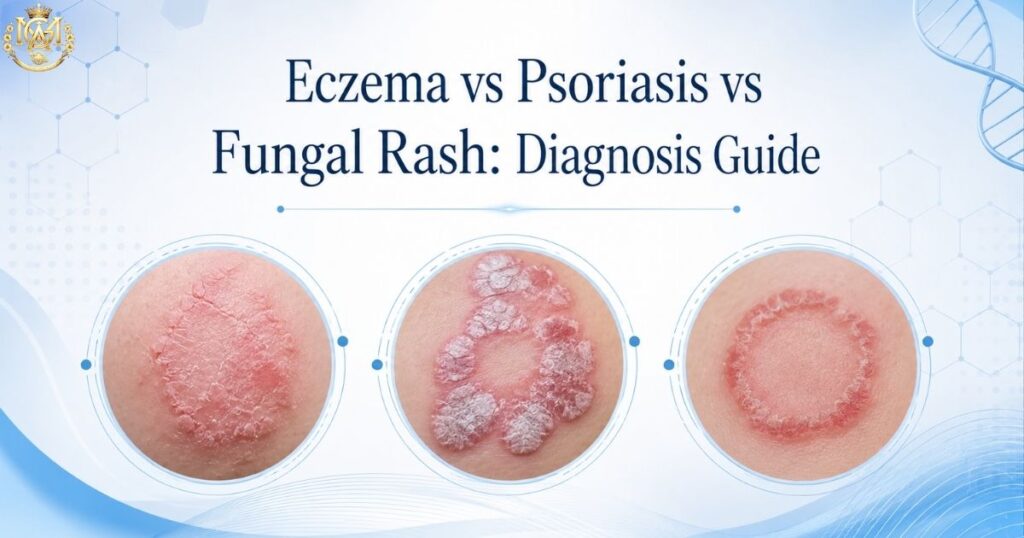
🩺 DIFFERENTIAL DIAGNOSIS MGA Clinical Education Series  ·  9 min read Eczema vs psoriasis vs fungal rash — three of the most commonly confused presentations in general practice — can look nearly identical at first glance. A patient walks into your clinic with an itchy, red, scaly rash on the elbows and knees… (rest of paragraph unchanged)A patient walks into your clinic with an itchy, red, scaly rash on the elbows and knees. Another presents with widespread, poorly-defined pale patches on the trunk after using an OTC cream. A third has a raised, silvery-scaled plaque on the extensor surface of the forearm that has been there for months. Three patients, three different conditions — but from a cursory clinical glance, they can look confusingly similar. Misdiagnosis between eczema (atopic dermatitis), psoriasis, and tinea (fungal rash) is remarkably common in general practice globally — and even more common in India, where steroid-modified tinea has become an epidemic masquerading as other conditions. This eczema vs psoriasis vs fungal rash guide walks through the key clinical differentiators that help a general physician reach the correct diagnosis quickly in the OPD, with a side-by-side comparison table and a simple decision framework that works in real-world practice. ⚡ Quick Differential Summary Eczema: Itch comes first → redness follows from scratching. Flexural surfaces (elbows, knees, neck). Lichenified skin, poor demarcation. Psoriasis: Well-demarcated plaques with silvery scale. Extensor surfaces (elbows, knees, shins, scalp). Systemic features possible (nail pitting, arthritis). Fungal (Tinea): Ring-shaped or atypical borders. Active border + central clearing (or pale centre if steroid-modified). KOH positive. Localized itch at border. Red flag: History of OTC cream use → suspect steroid-modified tinea mimicking eczema or psoriasis. 🔀 Why Eczema vs Psoriasis vs Fungal Rash Get Confused So Often All three conditions produce erythema (redness), scaling, and pruritus (itching). Each can present as a localized patch or become widespread, and each follows a chronic course. To an untrained eye — and occasionally even to a trained one — they appear nearly identical. The reason for confusion is structural: eczema and psoriasis are conditions of altered cutaneous immunity and barrier function; tinea is an infection. These arise from entirely different mechanisms — but the endpoint (inflammation, scaling, itch) is visually similar. Add to this the reality that in India, a fungal infection that has been treated with steroids (steroid-modified tinea) can look exactly like eczema or psoriasis, and misdiagnosis becomes not just common but expected. A machine-learning study published in Frontiers in Medicine (Ding et al., 2025, doi: 10.3389/fmed.2025.1667794) found that even with routine clinical data, eczema and psoriasis could not be distinguished with complete accuracy — illustrating that this differential is nuanced even with systematic approaches. The addition of a third condition (tinea) adds complexity but also opportunity: because tinea is fundamentally an infection, it responds to antifungals and has specific diagnostic tests (KOH), whereas eczema and psoriasis do not. 🧴 Eczema (Atopic Dermatitis): Key Clinical Features Classic phrase: “The itch that rashes” — not “the rash that itches.” This is the most clinically useful way to remember eczema’s hallmark. The itching comes first, often severe; scratching produces the visible rash and inflammation. 🔍 Eczema: Clinical Checklist Distribution: Flexural surfaces — antecubital fossa (crook of elbow), popliteal fossa (back of knee), neck, face, hands. In adults, often limited to hands or face. Children present with more widespread involvement. Appearance: Poorly demarcated (ill-defined borders). Lichenification (thickened, leather-like skin from chronic scratching). Erosions from scratching. Acute phase: vesicles and weeping. Chronic phase: dry, cracked, thickened skin. Itch character: Intense, often worse at night. Described as “maddening.” Scratch-itch cycle is central to perpetuation — the more you scratch, the more inflamed it becomes, the more it itches. Associated features: History of atopy (allergic asthma, allergic rhinitis, food allergy). Dry skin (xerosis) as baseline. May respond temporarily to steroids but relapses when stopped — if steroids alone are used without addressing barrier dysfunction. Response pattern: Responds well to emollients (moisturisers) + topical corticosteroids for acute flares + long-term barrier repair. Once correctly identified, it is predictable and manageable. 🏛️ Psoriasis: Key Clinical Features Classic phrase: “The rash that (sometimes) itches” — the plaque is present from the outset; itch is secondary and variable. An autoimmune condition in which the immune system attacks skin cells, causing rapid epidermal turnover. 🔍 Psoriasis: Clinical Checklist Distribution: Extensor surfaces — elbows, knees, shins, forearms, scalp, lower back, nails, ears. Psoriasis “loves bones” (bony prominences). Face and flexural areas are usually spared, which helps differentiate from eczema. Appearance: Well-demarcated plaques with a silvery or mica-like scale. The scale is often thick and adherent — when scraped off, pinpoint bleeding occurs (Auspitz sign). Sharp borders are characteristic. Lesions are thicker and more planar than eczema. Itch character: Variable — some patients report no itch at all; others report burning or soreness rather than intense itch. The plaque is visible and persistent; it does not improve simply from avoiding scratching. Associated features: Family history (positive in ~30% of patients). Nail involvement is common — pitting (tiny holes), onycholysis (separation from nail bed), subungual hyperkeratosis. Some patients have psoriatic arthritis (joint inflammation) — a key systemic clue not present in eczema or tinea. Response pattern: Chronic, cyclical (periods of remission and flare). Responds to topical steroids and vitamin-D analogues. Systemic triggers include stress, infections, certain medications (beta-blockers, lithium). Unlike tinea, psoriasis is never eradicated — it is managed long-term. 🍄 Fungal Rash (Tinea): Key Clinical Features Classic phrase: “Ring-shaped with a moving border.” The active border (periphery) is the site of active fungal invasion; the centre may clear (classic ringworm) or remain pale and poorly-defined if the patient has applied a steroid (steroid-modified tinea). 🔍 Fungal Rash (Tinea Corporis/Cruris/Faciei): Clinical Checklist Distribution: Any site — trunk, flexural and extensor surfaces equally affected. Tinea corporis affects the trunk and limbs, while tinea cruris involves the inguinal fold, pubic area, and inner thigh — the moist areas fungi favour. Tinea faciei, involving the face, is less common but increasing. Appearance: Classic:

Comparison infographic showing MD (university-based medicine), MS (university-based surgery), and DNB (NBEMS-based) postgraduate qualifications with key differences in training environment, assessment, and career pathways
Learning blogs

MD vs MS vs DNB: Which Postgraduate Path Should You Choose?

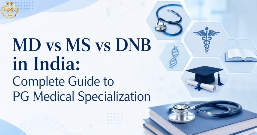
🎓 Complete Postgraduate Guide MD vs MS vs DNB in India: Complete Guide to PG Medical Specialization Regulatory equivalence ≠ training experience. This guide decodes the real differences, institutional quality factors, career pathways, and decision frameworks that shape your postgraduate choice. This MD vs MS vs DNB decision shapes everything from your daily training experience to your long-term career trajectory. The postgraduate medical pathway in India is shaped by a single dominant question: MD, MS, or DNB? While all three are recognised by the National Medical Commission (NMC) as “equivalent,” this equivalence exists only on paper. The actual training experience, clinical exposure, assessment rigour, institutional environment, and long-term career trajectory differ significantly — in ways that matter far more than the letters after your name. This guide moves beyond the surface-level “MD vs MS vs DNB comparison” found elsewhere. Instead, it decodes the clinical, structural, and regulatory differences that shape medical careers in India. By the end, you’ll understand not just what each qualification is, but how training institution quality, clinical exposure patterns, assessment rigour, and your specific long-term goals should drive your choice. ⚡ The Real Difference (At a Glance) MD University-based. Medical specialities. Structured mentorship. Assessment bias possible. Strong teaching college recognition. MS University-based. Surgical specialities. Procedure-heavy training. Operative skill-focused. Academic pathway strong. DNB NBEMS-based. Centrally assessed. Hospital-variable training. Standardised exit exam. Private/corporate pathways. 1️⃣ MD vs MS vs DNB: What These Qualifications Actually Mean MD: Doctor of Medicine An MD is a postgraduate degree awarded by universities recognised by the National Medical Commission, typically specialising in non-surgical disciplines. These include internal medicine, paediatrics, radiology, psychiatry, pathology, anaesthesia, and others. The degree is conferred after three years of hospital-based training, structured examinations, and submission of an academic dissertation. The Reality: MD training is designed around diagnostic reasoning, pharmacotherapy, and longitudinal patient management. An MD trainee spends substantial time in ward rounds, outpatient departments, diagnostic case discussions, and academic seminars. The exposure is breadth-oriented — seeing diverse presentations of a single disease domain — rather than depth-focused on procedural skill. MS: Master of Surgery An MS is the surgical counterpart to MD, awarded after three years of training in surgical disciplines: general surgery, orthopaedics, obstetrics and gynaecology, otolaryngology (ENT), and ophthalmology. The curriculum emphasises operative technique, perioperative decision-making, emergency surgical management, and procedural confidence. The Reality: MS trainees spend more time in operating theatres, emergency departments, and intensive care units than MD trainees. Their learning is anchored to procedural outcome — Was the patient positioned correctly? Was the dissection methodical? How would you manage this complication? This procedural focus shapes the entire clinical mindset. DNB: Diplomate of National Board A Diplomate of National Board (DNB) is a qualification awarded by the National Board of Examinations in Medical Sciences (NBEMS), an autonomous body under the Ministry of Health and Family Welfare. Unlike MD/MS, which are institution-specific degrees, the DNB is nationally uniform. Training occurs in NBEMS-accredited hospitals, which may be private, corporate, mission-based, or government institutions. The Reality: DNB is “standardised by design.” All candidates study the same broad curriculum, face identical exit examinations administered by external examiners, and receive a degree recognised across India uniformly. However, the training environment varies dramatically by institution — a DNB from a 600-bed private hospital in Mumbai may have vastly different clinical exposure than a DNB from a 150-bed mission hospital in a smaller city. 2️⃣ Eligibility and Admission Process Universal requirement: This MD vs MS vs DNB eligibility requirement is universal: all three pathways require an MBBS degree and qualification in the NEET-PG examination, held annually by the National Board of Examinations in Medical Sciences. Where Admission Differs MD/MS seats: Offered through central and state counselling in government and private medical colleges. Seat allocation is typically merit-based within state/national quotas. Preferred colleges (AIIMS, PGIMER, leading state colleges) fill quickly, and specialty preferences are limited by available seats. DNB seats: Filled through the same NEET-PG counselling but mapped to NBEMS-accredited hospitals. Total DNB seats are typically higher than MD/MS seats, offering greater flexibility for specialty and institution choice. Candidates often secure their preferred specialty through DNB when MD/MS seats are full. Critical point: Seat availability patterns mean your specialty choice may be determined by counselling luck rather than qualification type. A doctor who wanted Cardiology but couldn’t secure an MD/MS seat might pursue a DNB in Cardiology — a strategic choice, not a compromise. 3️⃣ MD vs MS vs DNB Training Duration and Structure Standard duration: MD vs MS vs DNB comparisons often start with duration — and here, MD, MS, and DNB programmes all typically span three years for core postgraduate training. However, the structure, rotations, and emphasis differ considerably. MD Training Structure Typically consists of: Daily ward rounds with faculty supervision Outpatient department rotations (managing clinic cases) Case presentations and journal clubs (weekly or bi-weekly) Rotations in related departments (e.g., cardiologists rotate through radiology, critical care) Research and dissertation on a chosen topic (mandatory) Internal assessments, mid-term exams, final university examinations MS Training Structure Typically consists of: Operating theatre rotations (assisting, observing, gradually taking operative roles) Ward rounds with emphasis on post-operative management Emergency department duties and on-call responsibilities Morbidity and mortality conferences (learning from complications) Rotations in related surgical departments (e.g., ortho residents train in trauma, emergency OT) Research and dissertation University examinations with heavy emphasis on operative cases and surgical scenarios DNB Training Structure Follows the same broad architecture as MD/MS but is institution-dependent: Clinical duties follow institutional protocols, not always standardised across India Academic components (seminars, journal clubs) vary by institution Research component is mandatory but structured differently than university MD/MS Exit examinations are NBEMS-conducted nationally (objective questions + practical) Centralised assessment means uniform standards but also variable institutional preparation 4️⃣ Clinical Training and Patient Exposure: Where Institution Matters Most This is where theory meets reality. The quality of your training experience depends more on the specific hospital, patient load, and faculty supervision than on whether you’re pursuing MD vs MS vs DNB. Government Teaching Hospital vs Private

Clinical comparison of classic tinea ringworm versus steroid-modified tinea in India — showing pale, poorly-defined lesions with blurred borders characteristic of OTC steroid cream misuse
Learning blogs

Steroid-Modified Tinea in India: The Silent Epidemic Every Doctor Should Understand

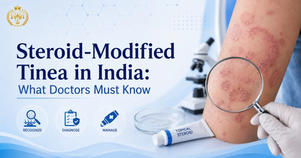
🩺 CLINICAL DERMATOLOGY MGA Clinical Education Series  ·  8 min read A patient walks into your OPD with a widespread, pale, mildly itchy skin rash on the trunk and groin. He has seen a pharmacist, used a cream, felt better for a week — and now it is back, larger and harder to define. This scenario plays out in dermatology OPDs and general physician clinics across India every single day. The culprit, in many of these cases, is not a new or unusual pathogen. It is an old fungal infection that has been allowed to change its face — because of a tube of OTC cream. This article explains what steroid-modified tinea is, why India is at the centre of a documented dermatology crisis, how these altered infections present clinically, why they are harder to clear, and what the published evidence says every treating doctor should do differently. 🔍 Quick Reference Condition: Steroid-modified tinea / Tinea incognito Cause: OTC topical steroid (or steroid–antifungal–antibiotic triple combination) applied to an undiagnosed dermatophyte infection Effect: Atypical clinical appearance, missed diagnosis, wider spread, treatment resistance Key Indian literature: IJDVL — Verma, Panda et al. 2021 (3-part series); IADVL–ITART Consensus 2020; Dutta, Rasul & Boro, IJDVL 2017 Doctor’s role: Clinical recognition, KOH confirmation, patient education, pharmacist communication 🔬 What Is Steroid-Modified Tinea (Tinea Incognito)? Tinea incognito — now more accurately called steroid-modified tinea in Indian clinical literature — refers to a dermatophyte infection whose typical clinical appearance has been altered by the application of a topical corticosteroid (or a calcineurin inhibitor). The term was first reported in 1968 by British dermatologists Dr Adrian Ive and Dr Ronnie Marks, who described patients with fungal infections masked by topical steroid use presenting as seborrheic dermatitis, eczema, or psoriasis-like eruptions. The mechanism is well understood: topical corticosteroids suppress local skin immunity — specifically, they downregulate antigen-presenting cell function, reduce IL-12 and Th1 activation, and shift the immune response toward a Th2 profile. The dermatophyte encounters a local immune environment that has been instructed to stand down. The inflammation that normally produces the classic raised, scaly, well-defined ring fades. The infection spreads laterally and sometimes deeper, but looks far less angry than it should. As published in Journal of Clinical Medicine (Zacharopoulou et al., 2024, PMC11172699), the more topical steroid that is applied, the more extensive and unrecognisable the fungal infection becomes. A critical nuance noted by Verma SB and colleagues in IJDVL (2021, doi: 10.25259/IJDVL_301_20) is that in the Indian epidemic, a strict “tinea incognito” — where the infection is completely unrecognisable — is less common than steroid-modified tinea, where an active border is often still visible despite morphological distortion. The IJDVL authors argue that “steroid-modified tinea” is therefore the more accurate term for the majority of Indian cases. 🏥 How an OTC Cream Habit Became a National Dermatology Concern India’s climate — high humidity, high ambient temperature across much of the country, and monsoon seasons that last months — creates conditions strongly favourable to dermatophyte growth and spread. Tinea corporis, tinea cruris, and tinea faciei are among the most common superficial infections seen in both urban and rural OPDs. In this environment, an itchy circular rash is a predictably common presentation. The problem is what happens next. Fixed-dose combination (FDC) creams containing a potent topical corticosteroid (typically clobetasol propionate or betamethasone dipropionate), an antifungal agent, and an antibacterial agent are widely available over the counter in India. Research published in the Indian Journal of Dermatology, Venereology and Leprology (Dutta, Rasul & Boro, IJDVL 2017) found that pharmacists were responsible for recommending these combination creams in the majority of tinea incognito cases — 78% of patients in that study were using products recommended by a pharmacist. Patients used these preparations for durations ranging from six weeks to twelve years without medical supervision. Why do patients keep using them? The steroid component produces rapid, visible relief within a few days. Itch decreases, redness fades. Patients interpret this as a cure. When the cream is stopped, the itch returns worse than before — because the fungus has been spreading unchecked beneath a suppressed immune response. The patient reaches for the tube again. The cycle repeats. A landmark 2021 three-part IJDVL series by Verma SB, Panda S, Nenoff P and colleagues described the situation as an “unprecedented epidemic-like scenario” of dermatophytosis in India, attributing the crisis directly to indiscriminate use of FDC corticosteroid creams and the emergence of a novel, partially terbinafine-resistant species — Trichophyton indotineae (formerly classified as T. mentagrophytes ITS genotype VIII) — which the WHO Fungal Priority Pathogens List (2022) has since acknowledged as a global concern. 📊 What the Published Research Documents • The prevalence of superficial dermatophytosis in different regions of India ranges from 36.6% to 78.4% — a range described by experts as constituting a national epidemic (topical antifungal-corticosteroid FDC review, Indian Journal of Pharmacology). • A cost-of-illness analysis published in Clinical and Experimental Dermatology (Jagadeesan et al., 2023) found that patients using topical steroids for dermatophytosis in India incur approximately 40% higher treatment costs than steroid-naive cases. • The IADVL Task Force Against Recalcitrant Tinea (ITART) Consensus 2020 noted a six- to seven-year pattern of unprecedented increase in recurrent, chronic, and steroid-modified dermatophytosis involving glabrous skin. • A study from Western Uttar Pradesh (Thakur et al., Journal of Dermatology and Cosmetology, 2018) conducted across 100 steroid-modified tinea cases identified lack of qualified dermatologists in rural areas as a compounding factor — a gap that directly increases irrational corticosteroid use. 👁️ Clinical Recognition: How Steroid-Modified Tinea Presents Differently The diagnostic challenge is that steroid-modified tinea no longer looks like textbook ringworm. The classic well-defined, raised, scaly, erythematous ring with central clearing is modified or absent. Recognising the altered patterns is the first clinical skill every doctor managing skin conditions in India needs to develop. Feature Classic Tinea (Untreated) Steroid-Modified Tinea Border Well-defined, raised, scaly Blurred, less elevated, less scaly Central clearing Present (classic ring shape) Often absent — study data shows 94.1% of steroid-modified

CPD accreditation for doctors in India — what it means, how it differs from CME and PG qualifications, and how to evaluate a credible program
Learning blogs

CPD Accreditation for Doctors in India: What It Means, Why It Matters and How to Choose a Program

Quick Answer CPD accreditation means a learning program has been assessed and approved by a recognised accrediting body — confirming its educational standards, faculty credibility, and clinical relevance. In India, CPD requirements are not uniformly mandated at the national level, but are increasingly tied to hospital credentialing, NMC’s evolving re-registration framework, and professional differentiation. A CPD-accredited fellowship certificate is different in nature from a university-awarded PG Diploma — understanding that difference is what helps you choose the right credential for your actual career goal. Most doctors encounter “CPD accredited” as a label on a course brochure without a clear sense of what that label actually guarantees — or who assigned it. Some programs carry accreditation from internationally recognised bodies with rigorous assessment criteria. Others carry badges from organisations that exist primarily to provide those badges. The label looks the same on a certificate; the substance behind it does not. This article explains what CPD accreditation is, how it differs from both CME and formal postgraduate qualifications like MD, MS, and PG Diplomas, what it unlocks practically in an Indian doctor’s career, and how to evaluate whether a CPD program is genuinely worth your time and fees. It is written for doctors who are already working — not for students trying to understand what CPD is in the abstract. CPD ≠ CME Different scope, different credential weight 3 credentials CPD certificate · PG Diploma · Fellowship — compared below NMC Framework for CPD in Indian medical practice is evolving What this article covers What CPD accreditation actually means — and what it doesn’t CPD vs CME — why the distinction matters in Indian practice What NMC’s evolving CPD framework means for practising doctors What CPD unlocks in practice — credentialing, career, and clinical authority CPD certificate vs Fellowship vs PG Diploma — which credential fits which goal How to evaluate whether a CPD program is credible Frequently asked questions What CPD Accreditation Actually Means — and What It Doesn’t Accreditation is an external quality mark. When a learning program is described as CPD accredited, it means a third-party body has reviewed the program’s curriculum, faculty credentials, assessment methods, and learning outcomes and confirmed that they meet defined educational standards. The accrediting body takes professional responsibility for that assessment. What accreditation does not mean: it does not automatically mean the program is government-approved, NMC-recognised, or equivalent to a postgraduate degree. These are separate categories with separate recognition criteria. A CPD-accredited program from a credible international body is a meaningful professional credential — but it is a different kind of credential from a university-awarded PG Diploma or an NMC-recognised MD/MS degree, and it should not be presented or understood as equivalent to either. The quality of accreditation varies significantly by the accrediting body. A program accredited by the CPD & Training Council (UK) — a body with defined international recognition in CPD standards — carries different weight than accreditation from a domestic body created specifically to issue certificates. Before enrolling in any CPD program, identify the accrediting body and verify independently that it is a recognised professional standards organisation, not a commercial certification provider. MGA’s accreditation: Fellowship programs at Medical Global Academy are accredited by the CPD & Training Council with international continuing medical education recognition. This is a professional CPD credential — not a university degree, not an NMC-recognised postgraduate qualification. The distinction matters for how you use the credential and what it demonstrates on a CV or in a hospital credentialing process. CPD vs CME — Why the Distinction Matters in Indian Practice CME (Continuing Medical Education) and CPD are used interchangeably in most Indian contexts — including by many medical associations. They are related but not identical, and understanding the difference helps you evaluate what a specific program is actually offering. Factor CME CPD Scope Primarily clinical knowledge updates Broader — clinical, professional, leadership, research, ethics Typical format Conferences, lectures, grand rounds Courses, workshops, teaching, mentorship, research, publications Learning goal Updated clinical knowledge Holistic professional development and career-long competency Credential output CME certificate / credit hours CPD certificate, fellowship credential, or structured learning record Relationship CME is a subset of CPD CPD includes CME and extends beyond it The practical implication: if a hospital credentialing process or employer asks for CPD records, attending an IMA conference generates CME credits that typically count. Completing a structured fellowship program generates CPD accreditation that carries deeper professional weight — it demonstrates sustained, assessed learning in a defined clinical area rather than attendance at a single event. What NMC’s Evolving CPD Framework Means for Practising Doctors India does not currently have a single, nationally uniform mandatory CPD requirement for all practising doctors. Requirements vary by State Medical Council — some states have introduced or are introducing renewal-linked CPD expectations; others have not formalised this. This landscape is actively evolving. The National Medical Commission has been developing a framework for continued medical education and professional development as part of broader regulatory modernisation. The direction of travel — toward structured CPD as part of ongoing medical registration maintenance — is consistent with international best practice in medical regulation. Doctors who have established CPD habits and documented learning records will be better positioned as this framework develops than those who begin scrambling when mandatory requirements arrive. Beyond national regulation, corporate hospitals and large healthcare networks in India have their own internal credentialing and re-credentialing processes that already incorporate CPD expectations — fellowship credentials, structured course completion, and documented specialty learning are factors in consultant appointment decisions at major private hospital groups independently of any state council requirement. Practical advice: Do not wait for CPD to become mandatory before building a record. The doctors who benefit most from structured CPD are those who treat it as an ongoing professional habit rather than a compliance task they manage at renewal deadlines. Verify current requirements directly with your State Medical Council — do not rely on generalised claims from any online source, including this one. What CPD Unlocks in

Doctor examining a patient representing recurrent fungal skin infections in India
Learning blogs

Fungal Skin Infections (Tinea) in India: Why They Keep Coming Back

Quick Answer Tinea (dermatophytosis) recurs so frequently in Indian patients for three converging reasons: the hot, humid climate creates near-ideal dermatophyte growth conditions year-round; widespread irrational use of topical steroid-antifungal combination products suppresses visible symptoms without eradicating the organism, selecting for resistant strains; and inadequate treatment duration means clinical clearance is achieved before mycological cure. Understanding these drivers — not just the presenting morphology — is what separates a GP who manages tinea effectively from one who keeps seeing the same patient back every monsoon. If you practise anywhere in India — in a city or a semi-rural setting, in a general OPD or a dedicated dermatology clinic — you have seen tinea. Almost certainly, you have seen the same patient return with it more than once. The question that should follow every relapsing tinea case is not “which antifungal should I prescribe this time” but “why did this come back.” India’s tinea burden has become a subject of specific concern in dermatology literature over the past decade, with Indian dermatologists documenting a pattern of recalcitrant, frequently recurrent dermatophytosis that appears to be more difficult to treat than the same presentations in previous decades. The reasons for this — climate, steroid misuse, treatment gaps, and emerging antifungal resistance — are discussed in this article at the level a general practitioner needs to understand to manage these cases more effectively. This is a two-part clinical discussion. Day 2 covers the full landscape of tinea in India — types, recurrence drivers, and general management principles. Day 3 goes deeper into the single most important driver of recurrence in the current Indian context: steroid-modified tinea and what it means for recognition, management, and outcome. That article is linked at the end of this one. Dermatophytes Primary causative organisms across all tinea types 3 drivers Climate + steroid misuse + inadequate treatment Trichophyton indotineae — the emerging resistant species of concern What this article covers The rising burden of fungal skin infections in India Common types — Tinea corporis, cruris, capitis and unguium Why recurrence is so common in Indian patients General principles of correct antifungal management Lifestyle and hygiene factors that worsen recurrence The hidden driver — topical steroid misuse (preview) Frequently asked questions The Rising Burden of Fungal Skin Infections in India Dermatophytosis — fungal infection of the skin, hair, and nails caused by dermatophyte fungi — is one of the most common infectious skin conditions globally. In India, it consistently ranks among the top presentations in both general practice and dermatology OPDs. The reported proportion of skin OPD attendances attributable to fungal infections in Indian studies typically ranges between 20–30%, with significant seasonal variation peaking during and immediately after the monsoon months (June–September). What has changed in recent years is not the frequency of presentation but the clinical behaviour. Indian dermatologists have documented a marked increase in presentations that are recurrent, widespread, and poorly responsive to standard antifungal regimens that previously worked reliably. Published case series and surveillance data from Indian dermatology centres have identified a specific organism — Trichophyton indotineae — as a newly characterised species that has emerged from the Indian subcontinent and shows elevated minimum inhibitory concentrations (MICs) to terbinafine, the most widely used oral antifungal for tinea in this country. The emergence of T. indotineae is not independent of clinical practice patterns — it is directly connected to the widespread misuse of topical corticosteroid-containing antifungal combination products and inadequate treatment courses, both of which have created selection pressure for resistant organisms. This is a public health-level problem with clinical-level consequences for every GP managing tinea. Common Types: Tinea Corporis, Cruris, Capitis and Unguium Tinea is named by anatomical site. The same causative organisms — primarily Trichophyton rubrum and Trichophyton mentagrophytes complex, increasingly T. indotineae in India — can infect different sites with different morphological presentations. Type Site Typical Morphology Indian Practice Notes Tinea corporis Trunk, limbs — any glabrous skin Annular or polycyclic plaques with active scaly border and central clearing Most commonly seen type; frequently steroid-modified — may lack classic ring morphology (covered in Day 3) Tinea cruris Groin, inner thighs, perineum Scaly erythematous plaques with defined advancing border; typically spares scrotal skin (unlike candidal intertrigo) More common in men; heat and occlusion major contributing factors; scrotum involvement suggests Candida or steroid-modified presentation Tinea capitis Scalp, eyebrows, eyelashes Patchy hair loss with scaling; grey-patch type (endothrix) or black-dot type; kerion (inflammatory, boggy mass) in severe cases Predominantly a paediatric presentation; oral antifungals required — topicals do not penetrate hair shaft; kerion warrants urgent management to prevent scarring alopecia Tinea unguium (onychomycosis) Nails — toenails more than fingernails Discolouration (yellow-white-brown), subungual hyperkeratosis, onycholysis, nail plate thickening and fragility Confirms fungal source — treat alongside active tinea corporis/cruris to prevent autoinoculation; longest treatment duration required of all tinea types Tinea pedis Interdigital web spaces, plantar surface Maceration, scaling, fissuring between toes; vesicular or hyperkeratotic plantar variant Often co-present with tinea unguium; common in those who wear closed shoes or walk barefoot in communal areas; a reservoir for tinea corporis/cruris autoinoculation Tinea faciei Face (non-beard) Annular or irregular erythematous patches; often atypical due to cosmetic product application and steroid use masking classic morphology Frequently misdiagnosed as eczema, rosacea, or seborrhoeic dermatitis; high index of suspicion required, especially in patients who apply fairness creams Why Recurrence Is So Common in Indian Patients Tinea recurrence is not a patient compliance problem, though compliance plays a role. It is predominantly a systems-level problem — a combination of environmental conditions, product availability, prescribing habits, and health system access that creates near-ideal conditions for dermatophytes to persist, reinfect, and develop resistance. 1. Climate and environmental load Dermatophytes are keratinophilic fungi that thrive at temperatures between 25–37°C and high ambient humidity. India’s tropical and subtropical climate provides these conditions through a significant portion of the year, and the monsoon months create conditions where sweating, skin maceration, and occlusive clothing converge. Unlike temperate climates where tinea is a seasonal problem, India provides near-continuous environmental conditions

General physician examining a patient representing acne management for general physicians in India
Learning blogs

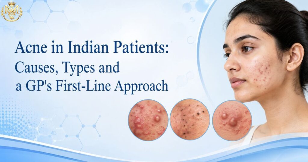
Acne in Indian Patients: Causes, Types and a GP’s First-Line Approach

Quick Answer Acne vulgaris is consistently the most common single dermatological presentation in Indian OPDs. First-line management for a GP spans three tiers based on severity: comedonal acne is primarily a topical retinoid case; mild-to-moderate papulopustular acne adds topical antibacterials; and moderate-to-severe or nodulocystic presentations are the clear trigger for dermatology referral rather than escalation in primary care. Indian skin’s higher baseline melanin content means post-inflammatory hyperpigmentation (PIH) is a clinical concern even in mild acne — this affects both how aggressively you treat active lesions and which agents carry additional pigmentary risk. Walk into any dermatology OPD in India and you’ll find acne. Walk into any general practice OPD and you’ll find it too — patients presenting not because they think it’s serious but because they’ve tried three or four over-the-counter products for six months and nothing is working, or because they’re developing scarring they didn’t expect, or because someone told them there’s a tablet that will fix it. Managing acne in a general practice setting doesn’t require dermatology training to do reasonably well at the first tier. It does require a clinical framework: knowing how to grade what you’re looking at, understanding what first-line management means and does not mean, recognising the patterns that need a dermatologist rather than an escalating course of antibiotics, and being specific about the pigmentation implications that matter more in Indian skin than in many published Western guidelines. This article covers all of that in a clinical format — for doctors managing acne in OPD, not for patients looking up their symptoms. Doctors who want to build structured dermatology competency beyond this first-line framework can explore MGA’s programmes on dermatology after MBBS. #1 Dermatology OPD presentation across age groups in India 3 tiers Grading-based management framework in this guide PIH The key Indian-skin consideration competitors omit What this article covers Why acne is the most common dermatology presentation in Indian OPDs Grading acne severity — comedonal to nodulocystic First-line management principles a general physician can apply Common prescribing mistakes to avoid Acne in Indian skin — pigmentation and scarring considerations When to refer to a dermatologist Frequently asked questions Why Acne Is the Most Common Dermatology Presentation in Indian OPDs Acne vulgaris affects the pilosebaceous unit — the follicle, its associated sebaceous gland, and the surrounding dermal tissue. The four pathophysiological processes driving it are well established: increased sebum production, abnormal follicular keratinisation (comedogenesis), colonisation by Cutibacterium acnes (formerly Propionibacterium acnes), and the resulting inflammatory cascade. Why it presents so commonly in Indian OPDs specifically comes down to a combination of factors: Age demographics: India has a large adolescent and young adult population — precisely the group in which androgen-driven sebum overproduction peaks. Most acne in this group is physiological in mechanism, not a sign of a hormonal disorder requiring investigation. Climate: Hot, humid conditions increase sweating and sebum secretion. Tropical acne — a more inflammatory form triggered by heat and humidity — is recognised as a distinct clinical variant, and overlaps substantially with the presentations seen in Indian summer months. Comedogenic cosmetics and topical product misuse: Oily hair oils and certain skin-lightening creams — both widely used across Indian demographics — are among the most common acneigenic topical products. Acne cosmetica and acne due to topical product misuse are more commonly encountered in Indian practice than most published guidelines address. Steroid misuse: The availability and widespread use of over-the-counter topical corticosteroid-containing products is a significant and growing contributor to steroid-modified acne and steroid acne presentations in Indian OPD — a pattern that is specifically documented in Indian dermatology literature (IJDVL). Grading Acne Severity: Comedonal to Nodulocystic Grading drives management. A GP who grades correctly at the first visit will make better treatment decisions, set more accurate patient expectations, and know more reliably when to refer. The most clinically practical grading framework for primary care is a three-tier system based on lesion type and extent. Grade Lesion Types Present Typical Distribution Management Implication Mild — Comedonal Open comedones (blackheads), closed comedones (whiteheads), minimal or no papules Nose, forehead, chin (T-zone) Topical retinoid — first-line; patience required (8–12 weeks for response) Mild to Moderate — Papulopustular Comedones + inflammatory papules and pustules; few to moderate in number Face ± upper trunk Topical retinoid + topical antibacterial; systemic antibiotics if topical inadequate after 6–8 weeks Moderate to Severe — Papulopustular / Nodular Numerous papules/pustules + nodules (>5 mm); possible early scarring Face, chest, back Refer to dermatologist; systemic antibiotics may bridge the gap while awaiting appointment Severe — Nodulocystic / Conglobate Multiple large nodules and cysts, interconnected sinuses possible, significant scarring Extensive face, trunk Dermatology referral without delay — oral isotretinoin territory; not a GP management case Lesion types: knowing what you’re looking at Non-inflammatory: Open comedones are blackheads — dilated follicular openings filled with oxidised keratin and sebum. Closed comedones are whiteheads — follicular plugs without a surface opening. Neither is inflamed, but both are precursors to inflammatory lesions if the follicle ruptures. Inflammatory: Papules are solid, elevated, inflamed lesions <5 mm. Pustules contain visible purulent material. Nodules are >5 mm, deeper, and more densely inflammatory. Cysts are fluctuant nodules — the defining feature of nodulocystic acne. Post-inflammatory: Not an acne lesion itself, but the aftermath — macules, PIH, and atrophic or hypertrophic scarring. In Indian skin, recognising PIH and early scarring as distinct from active lesions is important for both treatment sequencing and patient counselling. First-Line Management Principles a General Physician Can Apply The following principles are grounded in the Indian Dermatology guideline consensus and align with international acne management frameworks (American Academy of Dermatology, British Association of Dermatologists). No specific dosages are given here — these are prescribing-class principles, not a substitute for consulting a formulary or BNF equivalent. Principle 1: Address the comedone, not just the pustule The single most common first-line error in GP acne management is treating the visible inflammatory lesion while ignoring the comedonal base. Antibiotics — topical or oral — address the bacterial and inflammatory component

Scroll to Top