Medical Global Academy

Learning blogs

Doctors discussing the differences between a Fellowship in Oncology vs DM Medical Oncology career path
Learning blogs

Fellowship in Oncology vs DM Medical Oncology: Which Path Should You Choose?

📅 Updated September 2026 ⏰ 8 min read 🎓 MD Doctors Considering Oncology ⚡ Quick Answer Fellowship in Oncology vs DM Medical Oncology: what’s the real difference? DM Medical Oncology is a formal, NMC-recognised super-specialty degree — a three-year statutory qualification entered through NEET-SS, open to doctors holding MD/DNB in General Medicine, Paediatrics, or Radiation Oncology. A fellowship in oncology, including MGA’s, is a skill-building educational programme for eligible doctors — it builds oncology-focused knowledge but does not confer DM-level specialist status, statutory registration, or specialist recognition. These serve genuinely different purposes. Neither is a substitute for the other, and this guide explains exactly why — including a real regulatory development in 2026 that shows this distinction has practical consequences, not just academic ones. If you’re an MD doctor weighing a future in oncology, you’ve likely come across both terms — DM Medical Oncology and fellowship in oncology — sometimes used almost interchangeably in course listings and social media posts. They are not interchangeable. One is a regulated, statutory specialist pathway; the other is educational upskilling. Confusing the two can shape a career decision on inaccurate footing, which is exactly why this distinction deserves a clear, direct explanation before you choose either path. Fellowship in Oncology vs DM Medical Oncology: What Is the Difference? Factor Fellowship in Oncology DM Medical Oncology Entry point Eligible doctors as per the specific programme’s criteria MD/DNB in General Medicine, Paediatrics, or Radiation Oncology Purpose Additional oncology-focused knowledge and skills Formal super-specialty medical education Nature Skill-building educational programme Statutory, NMC-recognised specialist training Admission route Institutional selection process NEET-SS (national entrance) + centralised counselling Duration Typically 1–2 years, varies by programme 3 years Statutory specialist status Does not confer DM-equivalent status Part of the recognised super-specialty pathway What Is a Fellowship in Oncology? A fellowship in oncology is a structured educational programme designed to build oncology-focused clinical knowledge for doctors who are eligible under that specific programme’s criteria. Depending on the institution, this typically includes exposure to chemotherapy principles, systemic treatment approaches, multidisciplinary tumour board participation, and case-based learning in cancer diagnosis and management. The educational value here is real — doctors gain structured exposure to oncology decision-making they may not otherwise encounter. What a fellowship does not do is confer the statutory recognition, registration, or specialist title that comes with completing a super-specialty degree. It sits alongside formal training as a knowledge-building option, not a parallel route to the same destination. What Is DM Medical Oncology? DM Medical Oncology is a three-year, post-doctoral super-specialty degree recognised by the National Medical Commission. Entry is through NEET-SS (the national super-specialty entrance examination conducted by NBEMS), followed by centralised counselling. According to NBE’s own eligibility bulletin, the feeder qualifications for DM/DrNB Medical Oncology are an MD or DNB in General Medicine, an MD or DNB in Paediatrics, or an MD/DNB in Radiation Oncology — a doctor cannot enter this pathway directly from MBBS or from an unrelated MD. Training covers the systemic medical management of solid tumours and haematological cancers — chemotherapy, targeted therapy, immunotherapy, and hormonal therapy — alongside supportive and palliative care, and management of oncological emergencies. This is the pathway that leads to formal recognition as a medical oncologist in India. ℹ️ Eligibility criteria and seat availability for DM Medical Oncology can vary by institute and change between NEET-SS cycles. Always verify current-year requirements directly with NBEMS before making admission plans. Can an Oncology Fellowship Replace DM Medical Oncology? No. This deserves a direct answer rather than a hedge. A fellowship in oncology and DM Medical Oncology serve different purposes, have different eligibility requirements, and carry different professional positioning. A fellowship can help an eligible doctor build oncology-focused knowledge. It does not grant the same specialist qualification or statutory status as DM Medical Oncology, and it should never be represented as doing so. This isn’t just a technicality — it now has direct practical consequences. In April 2026, changes to eligibility rules under Ayushman Bharat PM-JAY, India’s national health insurance scheme, restricted empanelment for cancer treatment to oncologists holding NMC-recognised DM, MCh, or DrNB degrees, in several states. Reporting on the change described hundreds of experienced, fellowship-trained oncologists — including specialists trained at premier cancer centres — losing eligibility to treat patients under the scheme, regardless of years of clinical experience. ⚠️ Implementation has varied by state, and the policy has drawn pushback from affected doctors, with some ambiguity over whether NHA (which empanels hospitals) or NMC (which sets doctor qualification standards) holds final authority here. The details are still evolving — but the direction is clear: statutory super-specialty degrees and fellowship training are being treated as genuinely different tiers by regulators, not interchangeable credentials. Always verify current empanelment and registration requirements with the relevant authority before making career decisions based on this development. Fellowship in Oncology vs DM Medical Oncology: Which One Should You Choose? Choose Fellowship If Building Knowledge, Not Statutory Status You’re an eligible doctor seeking additional oncology-focused knowledge, exploring whether oncology genuinely fits your long-term interests, or want structured exposure without claiming — or needing — specialist equivalence. Choose DM Pathway If Pursuing Formal Specialist Recognition Your goal is formal, NMC-recognised medical oncology super-specialist status. You hold (or plan to complete) the required MD/DNB base qualification, and you’re prepared for the competitive NEET-SS admission process and the full three-year training commitment. A simple decision rule: if your current qualification is an eligible MD/DNB and your goal is formal specialist status, DM Medical Oncology through NEET-SS is the relevant route. If you’re looking to build oncology-focused knowledge and skills without pursuing that statutory route right now, a fellowship serves a genuinely different, complementary purpose — not a shortcut to the same outcome. Frequently Asked Questions Is a fellowship in oncology equivalent to DM Medical Oncology?▼ No. DM Medical Oncology is a statutory, NMC-recognised super-specialty degree. A fellowship in oncology is a skill-building educational programme. They differ in eligibility, recognition, admission process, and professional positioning — a fellowship does not confer DM-equivalent
Medical student evaluating options to find the easiest medical specialization to get in India
Learning blogs

Easiest Medical Specialization to Get India (And the Hardest Ones to Crack)

📅 Updated September 2026 ⏰ 8 min read 🎓 NEET PG Aspirants 🌎 Specialization Cluster — Blog 7 ⚡ Quick Answer Which medical specializations are easiest or hardest to get after MBBS in India? Competition for a NEET PG branch depends on how many seats exist nationally relative to how many aspirants want them — not on the specialty’s clinical difficulty. Branches like Dermatology, Radiodiagnosis, and General Surgery in premier institutes traditionally see higher competition, while General Medicine, Community Medicine, and Pathology often offer comparatively wider availability, particularly outside top-tier colleges. Exact cutoffs and seat numbers change every admission cycle — this guide explains the structural reasons behind these patterns rather than quoting figures that go stale within months. Every NEET PG aspirant eventually hears the same informal rankings — “Dermatology is impossible to get,” “Community Medicine is easy,” and so on. There’s real substance behind these labels, but they’re often repeated without anyone explaining why, which leaves aspirants calibrating their expectations against vague folklore rather than an actual understanding of how competition works. This guide explains the structural reasons some branches see more competition than others, which specialties traditionally fall on each side of that line, and — just as importantly — why “easier to get” has nothing to do with a specialty’s actual worth as a career. What Makes a NEET PG Branch “Hard” or “Easy” to Get A branch becomes competitive for one straightforward reason: more aspirants want it than there are seats to go around, relative to the applicant pool’s rank distribution. That’s it — competition level says nothing about how intellectually demanding the specialty is, how many years of training it requires, or how satisfying a career in it turns out to be. This also means “hard” and “easy” aren’t fixed facts — they’re a snapshot of demand and supply that shifts, sometimes meaningfully, from one admission cycle to the next as aspirant preferences, seat counts, and counselling dynamics change. A branch seen as difficult to secure a few years ago can look different today, and vice versa. Structural Factors: Seat Count, Popularity, and Category Dynamics Three structural factors drive most of the variation you’ll hear about. Total seat count Some branches simply have far fewer postgraduate seats nationally than others, spread across a smaller number of institutions. A branch with a naturally limited seat pool will look more competitive purely on supply, regardless of how many people actually want it. Popularity relative to seats Certain branches attract disproportionate aspirant interest — often driven by perceived lifestyle, income potential, or procedural appeal — independent of how many seats actually exist. When popularity outpaces seat count, competition rises sharply even in branches that aren’t structurally scarce. Category and quota dynamics Reservation categories and state-domicile quotas mean the same seat can be genuinely competitive for one candidate and comparatively more accessible for another, even within the identical college and specialty. Institution tier matters too — a branch in a premier government institute is virtually always more competitive than the identical branch in a lesser-known private college, regardless of the specialty’s general reputation. ⚠️ Because these factors interact and shift yearly, this article deliberately avoids quoting specific cutoff ranks, percentiles, or seat numbers — any figure here would be stale by the time you read it. Always check the current year’s official NBEMS counselling data for verified numbers. Branches With Traditionally Higher Competition Based on longstanding aspirant demand patterns rather than any single year’s data, a few branches are consistently discussed as more competitive: Dermatology Strong outpatient-based lifestyle appeal and growing cosmetic dermatology demand keep aspirant interest consistently high relative to the limited number of seats available. Radiodiagnosis Perceived favourable work-life balance combined with strong long-term earning potential has made this one of the more sought-after physician-track branches for years. General Surgery (Premier Institutes) While General Surgery itself has a reasonably large national seat pool, seats specifically at premier government institutions remain intensely competitive. Orthopaedics Combines strong private-practice earning potential with a procedural, technique-driven appeal that consistently draws high aspirant interest nationally. Emergency Medicine and select branches like ENT and Ophthalmology also see above-average competition in certain states and institution types, though this varies more by region than the branches above. Branches With Traditionally Better Availability On the other side, several branches generally offer more realistic seat availability for a broader range of ranks: General Medicine (Beyond Premier Colleges) A genuinely large national seat pool, particularly outside the most sought-after institutions, makes this one of the more consistently accessible MD branches. Community Medicine (PSM) Lower aspirant demand relative to its seat count, despite genuine importance in public health, epidemiology, and health administration career paths. Pathology Comparatively wider availability in many regions, offering strong diagnostic and academic career depth without matching the aspirant demand seen in clinical branches. Anaesthesiology (Non-Metro Colleges) Generally more accessible outside major metro institutions, despite strong demand and career stability once qualified. Forensic Medicine and pre-clinical academic branches like Physiology and Biochemistry also tend to offer wider availability, reflecting their lower profile among aspirants rather than any lack of academic or career value. “Easier to Get” Doesn’t Mean “Lower Value” — Why That’s a Myth This is worth stating plainly: competition level reflects aspirant demand at a point in time, not a specialty’s clinical importance, career stability, or growth potential. Treating “easier to get” branches as consolation prizes does both the specialty and the doctor choosing it a disservice. Community Medicine graduates go on to shape public health policy, lead epidemiological responses, and hold senior administrative roles that clinical branches rarely offer a direct path into. Pathology sits at the diagnostic center of every hospital, with genuine private lab and academic research opportunities. General Medicine remains one of the most versatile, consistently in-demand qualifications across every practice setting in the country — hospital-employed, private consultant, or rural general practice. If a specialty’s day-to-day work genuinely interests you, lower competition is a practical advantage, not a compromise. The mistake isn’t choosing a less competitive branch

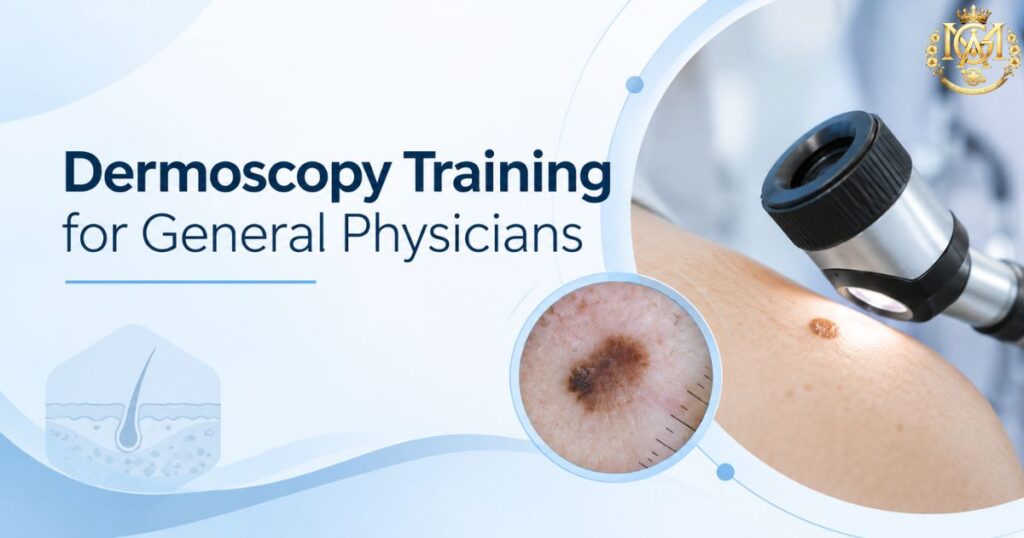
General physician receiving dermoscopy training to improve non-invasive skin diagnosis
Learning blogs

Dermoscopy for General Physicians: Skin Diagnosis, Differential Diagnosis & When to Refer

📅 Updated September 2026 ⏰ 8 min read 🩺 General Physicians & MBBS 🌎 Dermatology Cluster — Day 8 ⚡ Quick Answer Why should general physicians learn basic dermoscopy? It’s rarely taught during MBBS. Yet it meaningfully improves diagnostic confidence in exactly the situations GPs face daily — distinguishing fungal from eczema or psoriasis, and recognising when a pigmented lesion needs urgent referral. It doesn’t replace clinical judgement or specialist referral — it adds a second layer of information before you decide to treat or refer. Ask most MBBS graduates what a dermatoscope is, and you’ll often get a vague answer — “something dermatologists use to look at skin.” That vagueness reflects a real gap. Dermoscopy is rarely part of the standard MBBS curriculum. Most general physicians go through an entire career without seeing it — despite skin complaints being one of the most common reasons patients walk into a general OPD.. This is worth addressing directly, because dermoscopy isn’t an obscure specialist tool — it’s a practical skill that sharpens exactly the kind of differential diagnosis decisions GPs make constantly. Skin presentations are among the most frequent reasons patients seek a general consultation. Yet the diagnostic toolkit most GPs bring to that consultation hasn’t changed much. It still relies mainly on visual inspection and history-taking.. Dermoscopy fills a genuine gap in that toolkit, without requiring a full dermatology residency to gain real value from it. This guide covers what dermoscopy is and what it helps a non-specialist recognise. It also covers its role in differential diagnosis, skin cancer awareness, and what structured learning looks like.. What Is Dermoscopy and Why It’s Underused Outside Dermatology Dermoscopy uses a handheld device called a dermatoscope. It combines magnification with a light source — either polarised or in contact with the skin via an interface fluid. This lets you visualise structures beneath the skin’s surface that aren’t visible to the naked eye.. This includes pigment patterns, vascular arrangements, and follicular openings that carry real diagnostic information. The tool itself isn’t complex or expensive to use, but three factors keep it largely confined to dermatology practice. First, it simply isn’t part of the standard MBBS curriculum at most institutions, so exposure typically only happens during dermatology postgraduate training. Second, interpreting what you see through a dermatoscope takes structured learning — pattern recognition here isn’t intuitive the way basic clinical examination is.Third, many doctors assume dermoscopy is purely a cancer-screening tool for specialists. That misconception undersells its everyday value in routine differential diagnosis. This is somewhat unusual compared to other diagnostic aids in medicine. A stethoscope, an otoscope, or a basic ophthalmoscope are all standard equipment GPs are trained to use during MBBS, despite requiring their own learning curve. Dermoscopy sits in a strange gap — genuinely useful, not prohibitively difficult to learn at a basic level, yet rarely introduced as part of core training. That gap is exactly what makes it worth actively seeking out rather than waiting to encounter it by chance. What Dermoscopy Can Help a Non-Specialist Recognise Even at a basic, non-expert level, dermoscopy adds genuinely useful information to a standard clinical exam. Vascular Pattern Recognition Distinguishing the type and arrangement of blood vessels visible in a lesion — a detail invisible to the naked eye but often characteristic of specific conditions. Scale & Surface Structure Identifying scale patterns, follicular plugging, and comedo-like openings that help differentiate inflammatory from infective skin conditions. Pigment Network Assessment Seeing whether a pigmented lesion’s structure looks regular and symmetric, or irregular enough to warrant a closer specialist look. Hair & Follicular Changes Recognising characteristic hair shaft changes that support a fungal diagnosis in hair-bearing skin, alongside standard KOH testing. The value here isn’t that dermoscopy replaces clinical judgement — it’s that it adds a second, objective layer of information before you decide whether to treat, watch, or refer. That’s a meaningfully different position than relying on morphology and history alone. Dermoscopy in Differential Diagnosis: Tinea, Eczema, Psoriasis This is where dermoscopy earns its keep in daily general practice. Our earlier guide on eczema vs psoriasis vs fungal rash covers the clinical differentiators between these three commonly confused conditions — dermoscopy adds a further layer of objective evidence on top of that clinical picture. Condition What Dermoscopy Adds Tinea (Fungal) Characteristic hair shaft changes in affected hair-bearing skin, supporting the diagnosis alongside KOH testing Psoriasis A fairly regular, evenly distributed vascular pattern against a light background, with diffuse white scale Eczema Less specific findings — often patchy vessel distribution with a yellowish scale or serous crust pattern, distinct from psoriasis’s regularity This is particularly valuable in exactly the scenario our differential diagnosis guide flags as the trickiest: steroid-modified presentations, where a fungal infection has been altered by inappropriate steroid cream use and no longer shows classic clinical features. In these atypical cases, combining dermoscopy findings with a KOH scraping provides meaningfully more diagnostic confidence than relying on clinical morphology alone. Dermoscopy for Early Skin Cancer Screening Awareness Dermoscopy is well established in dermatology as improving diagnostic accuracy for pigmented lesion assessment compared to naked-eye examination alone. For general physicians, the relevant takeaway isn’t to become a diagnostician for skin cancer — it’s to develop enough pattern awareness to recognise when a pigmented lesion looks atypical and needs prompt specialist referral, rather than a “watch and wait” approach. Broad awareness points worth building familiarity with include asymmetry in a lesion’s shape, irregular or poorly defined borders, variation in colour within a single lesion, and any lesion that’s changing in size, shape, or appearance over time. None of these features alone confirm malignancy, and none of them should be used as a standalone diagnostic checklist by a non-specialist — they’re a screening lens for deciding who needs a dermatologist’s assessment sooner rather than later. ⚠️ Dermoscopy at the GP level is a triage and referral tool, not a diagnostic endpoint. Any lesion that looks atypical on dermoscopic examination still requires dermatology assessment and, where indicated, biopsy —

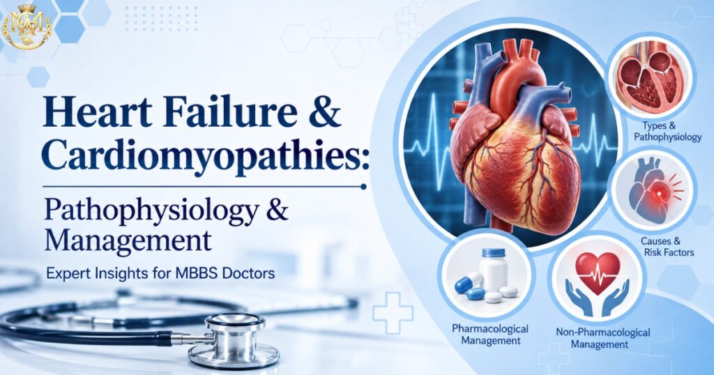
Cardiologist reviewing an echocardiogram representing pathophysiology of heart failure and cardiomyopathies
Learning blogs

Heart Failure and Cardiomyopathies: Understanding the Heart’s Struggle to Keep Up

Cardiovascular Health A clear guide to pathophysiology, neurohormonal remodeling, genetics, and modern management — from first-line medication to transplant. In This Guide Pathophysiology of Heart Failure Management of Heart Failure Cardiomyopathies: The Four Major Types A Condition That Rewards Early, Coordinated Care Heart failure isn’t a single disease — it’s the endpoint of many different paths the heart’s muscle, valves, or rhythm can take when something goes wrong. Understanding heart failure and cardiomyopathies together matters, because cardiomyopathies are among the most important underlying causes of heart failure itself. At its core, heart failure means the heart can no longer pump enough blood to meet the body’s needs, or can only do so at abnormally high filling pressures. Cardiomyopathies — diseases that begin in the heart muscle itself — explain why treatment for heart failure and cardiomyopathies today looks so different from a decade ago. For doctors looking to build deeper expertise in this area, MGA’s Fellowship and PG Diploma programmes offer structured, CPD-accredited training across cardiovascular and related specialties. Pathophysiology of Heart Failure Systolic vs. Diastolic Dysfunction Every failing heart struggles with one of two basic problems, sometimes both at once. In systolic dysfunction, the heart muscle contracts weakly, so it can’t squeeze out enough blood with each beat — this is what most people picture when they hear “heart failure,” and it’s measured by a reduced ejection fraction. In diastolic dysfunction, the muscle contracts normally but has become stiff, so the chambers can’t relax and fill properly between beats, even though the pump itself is still strong. This systolic-versus-diastolic distinction is central to understanding heart failure and cardiomyopathies together, since it determines which treatments actually help. Neurohormonal Regulation and Cardiac Remodeling When the heart first starts struggling, the body tries to help. The sympathetic nervous system raises heart rate and contractile force; the renin-angiotensin-aldosterone system (RAAS) retains salt and water to boost circulating volume. In the short term, these responses genuinely support circulation. Sustained over months and years, though, they become the disease’s own engine — constant neurohormonal activation drives the heart to enlarge, thicken, and stiffen in a process called remodeling, which further impairs pump function and closes a vicious cycle. Why this matters clinically: Nearly every major heart failure medication class exists specifically to interrupt this neurohormonal cycle — which is exactly why understanding remodeling comes before understanding treatment, not after. Genetic Predispositions A meaningful share of heart failure, especially in younger patients, traces back to inherited mutations in the genes that build the heart’s structural proteins — the sarcomere, the cytoskeleton, the ion channels that keep the heartbeat coordinated. Recognizing a genetic cause changes the conversation from “what happened to this heart” to “who else in the family might be at risk,” which is why family history deserves as much attention as any laboratory test. A heart failure diagnosis with no obvious cause — no prior heart attack, no long-standing hypertension, no valve disease — is often exactly the situation where a genetic origin becomes worth investigating early, rather than as a last resort. The NIH’s overview of cardiomyopathy genetics covers this diagnostic pathway in more depth. Management of Heart Failure Pharmacological Therapy Four drug classes now form the backbone of treatment for heart failure with reduced ejection fraction, often introduced together rather than one at a time. RAAS inhibitors — ACE inhibitors, ARBs, or the newer angiotensin receptor–neprilysin inhibitors — blunt the very hormonal drive that fuels remodeling. Beta-blockers calm chronic sympathetic overactivation. SGLT2 inhibitors, originally developed for diabetes, have earned a place across nearly the full spectrum of heart failure, reducing hospitalizations regardless of a patient’s blood sugar status. Diuretics remain essential for day-to-day symptom relief, easing congestion even though they don’t extend life on their own. The modern approach favors starting several of these classes early and in parallel, rather than the older step-by-step model, because the sooner remodeling is interrupted, the more function tends to be preserved. Advanced Treatments: CRT and ICDs For patients whose hearts also struggle electrically, resynchronization therapy can help — a specialized pacemaker restores coordinated contraction when the heart’s chambers have fallen out of step with each other. A related but distinct device, the implantable cardioverter-defibrillator, exists purely to prevent sudden death by recognizing and correcting dangerous rhythms before they become fatal. Many patients with more advanced heart failure qualify for both. Advanced Heart Failure Therapies When medication and devices are no longer enough, mechanical and surgical options come into view. A left ventricular assist device takes over much of the heart’s pumping work mechanically, either as a bridge while awaiting transplant or, increasingly, as a long-term therapy in its own right. Heart transplantation remains the definitive option for eligible patients with end-stage disease, offering a chance at meaningfully longer, fuller life when nothing less invasive can keep pace with the heart’s decline. A recent narrative review of emerging heart failure therapies covers where this field is heading next. Cardiomyopathies The Four Major Types Hypertrophic cardiomyopathy thickens the heart muscle, often unevenly, and can obstruct blood flow out of the heart while raising the risk of dangerous rhythms — it’s a leading cause of sudden cardiac death in young athletes. Dilated cardiomyopathy stretches and weakens the ventricles, the most common structural pathway into systolic heart failure. Restrictive cardiomyopathy, the rarest of the group, stiffens the heart muscle without necessarily enlarging it, so the chambers resist filling even when contraction still works. Arrhythmogenic right ventricular cardiomyopathy replaces normal heart muscle, usually on the right side, with fatty and fibrous tissue, creating fertile ground for dangerous arrhythmias, often in otherwise healthy-looking young people — a pattern examined closely in this review of arrhythmogenic cardiomyopathy management. Genetic Testing and Counselling Because so many cardiomyopathies run in families, genetic testing has become a standard part of the diagnostic pathway once one is suspected or confirmed. A positive result doesn’t just inform the patient in front of you — it opens the door to cascade screening for first-degree relatives, some of whom

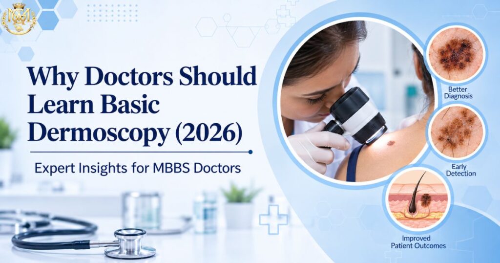
Graphic illustrating why doctors should learn basic dermoscopy, showing a physician examining a patient alongside magnified skin lesions
Learning blogs

Why Every General Physician Should Learn Basic Dermoscopy

📅 Updated September 2026 ⏰ 8 min read 🩺 General Physicians & MBBS 🌎 Dermatology Cluster — Day 8 ⚡ Quick Answer Why should general physicians learn basic dermoscopy? Dermoscopy is a non-invasive technique using a handheld magnifying device to see skin structures below the surface that aren’t visible to the naked eye. It’s rarely taught during MBBS, yet it meaningfully improves diagnostic confidence in exactly the situations GPs face daily — distinguishing fungal infections from eczema or psoriasis, and recognising when a pigmented lesion looks atypical enough to warrant urgent referral. It doesn’t replace clinical judgement or specialist referral — it adds a second layer of information before you decide to treat or refer. Ask most MBBS graduates what a dermatoscope is, and you’ll often get a vague answer — “something dermatologists use to look at skin.” That vagueness reflects a real gap: structured dermoscopy training for general physicians remains rare, and most GPs go through an entire career without direct exposure to the skill, despite skin complaints being among the most common reasons patients walk into a general OPD. TThis is worth addressing directly. Dermoscopy isn’t an obscure specialist tool — it’s a practical skill that sharpens the differential diagnosis decisions GPs make constantly. Skin presentations are among the most frequent reasons patients seek a general consultation. Yet the diagnostic toolkit most GPs bring to that consultation hasn’t changed much beyond visual inspection and history-taking. Dermoscopy fills a genuine gap in that toolkit, without requiring a full dermatology residency to gain real value from it.. This guide covers what dermoscopy actually is, what it can help a non-specialist recognise, where it fits into common differential diagnosis dilemmas, its role in early skin cancer awareness, and what structured learning actually looks like. What Is Dermoscopy, and Why Do So Few General Physicians Get Trained In It?y Dermoscopy uses a handheld device called a dermatoscope. It combines magnification with a light source, either polarised or in direct contact with the skin using an interface fluid. This lets doctors visualise structures beneath the skin’s surface that aren’t visible to the naked eye. This includes pigment patterns, vascular arrangements, and follicular openings that carry real diagnostic information. The tool itself isn’t complex or expensive to use, but three factors keep it largely confined to dermatology practice. First, it simply isn’t part of the standard MBBS curriculum at most institutions, so exposure typically only happens during dermatology postgraduate training. Second, interpreting what you see through a dermatoscope takes structured learning — pattern recognition here isn’t intuitive the way basic clinical examination is. Third, there’s a common misconception that dermoscopy is purely a cancer-screening tool for specialists, which undersells its everyday value in routine differential diagnosis. This is somewhat unusual compared to other diagnostic aids in medicine. A stethoscope, an otoscope, or a basic ophthalmoscope are all standard equipment GPs learn to use during MBBS. Each of these tools has its own learning curve too. Dermoscopy sits in a strange gap — genuinely useful, not prohibitively difficult to learn at a basic level. Yet core curricula rarely introduce it. That gap is exactly what makes it worth actively seeking out rather than waiting to encounter it by chance What Dermoscopy Can Help a Non-Specialist Recognise Even at a basic, non-expert level, dermoscopy adds genuinely useful information to a standard clinical exam. Vascular Pattern Recognition Distinguishing the type and arrangement of blood vessels visible in a lesion — a detail invisible to the naked eye but often characteristic of specific conditions. Scale & Surface Structure Identifying scale patterns, follicular plugging, and comedo-like openings that help differentiate inflammatory from infective skin conditions. Pigment Network Assessment Seeing whether a pigmented lesion’s structure looks regular and symmetric, or irregular enough to warrant a closer specialist look. Hair & Follicular Changes Recognising characteristic hair shaft changes that support a fungal diagnosis in hair-bearing skin, alongside standard KOH testing. The value here isn’t that dermoscopy replaces clinical judgement — it’s that it adds a second, objective layer of information before you decide whether to treat, watch, or refer. That’s a meaningfully different position than relying on morphology and history alone. Dermoscopy in Differential Diagnosis: Tinea, Eczema, Psoriasis This is where dermoscopy earns its keep in daily general practice. Our earlier guide on eczema vs psoriasis vs fungal rash covers the clinical differentiators between these three commonly confused conditions — dermoscopy adds a further layer of objective evidence on top of that clinical picture. Condition What Dermoscopy Adds Tinea (Fungal) Characteristic hair shaft changes in affected hair-bearing skin, supporting the diagnosis alongside KOH testing Psoriasis A fairly regular, evenly distributed vascular pattern against a light background, with diffuse white scale Eczema Less specific findings — often patchy vessel distribution with a yellowish scale or serous crust pattern, distinct from psoriasis’s regularity This is particularly valuable in exactly the scenario our differential diagnosis guide flags as the trickiest: steroid-modified presentations. In these presentations, inappropriate steroid cream use has altered a fungal infection that no longer shows classic clinical features. Combining dermoscopy findings with a KOH scraping adds meaningfully more diagnostic confidence in these atypical cases than clinical morphology alone.” Early Skin Cancer Screening: What General Physicians Should Know About Dermoscopy Dermoscopy is well established in dermatology as improving diagnostic accuracy for pigmented lesion assessment compared to naked-eye examination alone — a finding echoed in a systematic review of dermoscopy use for melanoma detection and triage in primary care settings. For general physicians, the relevant takeaway isn’t to become a diagnostician for skin cancer — it’s to develop enough pattern awareness to recognise when a pigmented lesion looks atypical and needs prompt specialist referral, rather than a “watch and wait” approach. Broad awareness points worth building familiarity with include asymmetry in a lesion’s shape and irregular or poorly defined borders. Others include colour variation within a single lesion, and any lesion changing in size, shape, or appearance over time. None of these features alone confirm malignancy. A non-specialist shouldn’t rely on any

Surgeons in an operating room representing MS surgical specialization training and lifestyle
Learning blogs

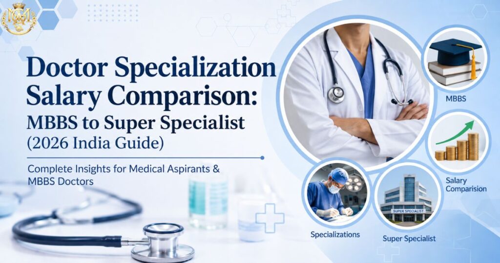
Doctor Specialization Salary Comparison: MBBS to Super Specialist (2026 India Guide

📅 Updated September 2026 ⏰ 11 min read 🎓 MBBS to Super Specialist 🌎 Specialization × Salary Cluster ⚡ Quick Answer How does doctor salary in India change from MBBS to super specialist? This doctor specialization salary comparison shows how pay scales in stages by qualification level, not just experience. An MBBS fresher typically earns ₹40,000–₹90,000 a month. An MD/MS consultant moves into the ₹1 lakh–₹3.3 lakh range depending on seniority. An established DM/MCh super specialist can earn ₹4.2 lakh–₹16.7 lakh a month, with senior national-level experts reaching ₹6.7 lakh–₹25 lakh. Every figure below is pulled directly from the same reference data used across our complete doctor salary guide. That keeps the numbers here fully consistent with it. Why This Doctor Specialization Salary Comparison Starts With Qualification Level This doctor specialization salary comparison begins with a common question doctors ask: “which specialty pays the most?” But the more useful question is how income actually changes as you move up the qualification ladder. The honest answer is that salary in Indian medicine scales in fairly distinct stages. Those stages are MBBS, then MD or MS, then DM/MCh for those who go on to super-specialise. Each step doesn’t just add a bit more pay — it typically unlocks a meaningfully different earning band. This guide walks through the full ladder using the same reference data behind our complete doctor salary guide. Whether you’re comparing a single stage or the entire career arc, the numbers stay consistent. You won’t find contradictions anywhere else on the site. If you’re still deciding which specialty to pursue, look at this ladder first. It matters more than any single specialty’s headline number. Two doctors in very different fields — a General Medicine physician and an Orthopaedic surgeon — can earn similar incomes. That’s simply because they’re at the same qualification stage. The specialty-specific differences described in our highest paying medical specializations guide matter too. But they become far more meaningful once you’ve factored in where you sit on this broader ladder. How Doctor Salaries Scale With Qualification Level Three things drive where you sit on the salary ladder at any point. The first is your qualification level — MBBS, MD/MS, or DM/MCh. The second is your years of experience within that level. The third is your practice setting — government, private hospital, or independent practice. Qualification level sets the band you’re in. Experience moves you up within that band. Practice setting determines where within the range you actually land. What’s worth understanding upfront is that these bands overlap. A highly experienced MBBS doctor without a postgraduate degree can, in some cases, out-earn a newly qualified MD consultant. But the ceiling for further growth is structurally different. Postgraduate and super-specialist qualifications don’t just pay more on average; they unlock significantly higher ceilings as experience accumulates. It’s also worth being clear about what these figures represent. Each range reflects typical monthly pay across government postings, private hospitals, and independent or consulting-based practice. It isn’t a single fixed number. Where you land within a given range depends heavily on which setting you’re in. It also depends on your specific city and institution. Two doctors at the identical qualification stage can sit at opposite ends of the same range for exactly this reason. MBBS-Level Earnings (Internship to 10+ Years, No PG) Before any postgraduate qualification, earnings still grow meaningfully with experience. That growth just sits within a narrower band than what postgraduate training eventually unlocks. Career Stage Monthly Range Internship ₹15,000 – ₹35,000 MBBS Fresher (0–2 yr) ₹40,000 – ₹90,000 MBBS 3–5 years ₹80,000 – ₹1.5 lakh MBBS 5–10 years ₹1 lakh – ₹2 lakh MBBS Senior (10+ yr, no PG) ₹1.5 lakh – ₹2.2 lakh Internship is a training stipend, not a full salary. It reflects your status as a student completing mandatory clinical rotations rather than an independently practising doctor. Once you cross into fresher territory, most doctors take roles like Medical Officer, RMO, or Duty Doctor. These roles are stepping stones while they decide their next move. The jump from fresher to the 3–5 year mark typically reflects growing clinical confidence. In many cases, it also marks a shift from purely rotational duty roles into more defined, better-compensated positions. By the 5–10 year mark without a postgraduate qualification, earnings tend to plateau. This is precisely the point at which MD, MS, or DNB training becomes the clearest lever for continued income growth. It’s not just about clinical growth anymore. The gap between MBBS Senior (₹1.5 lakh–₹2.2 lakh) and MD Senior Consultant (₹2.1 lakh–₹3.3 lakh) illustrates this well. A postgraduate qualification doesn’t just add incremental pay — it opens an entirely different ceiling for the years that follow. MD Salary Progression (Junior to Senior Consultant) Career Stage Monthly Range MD Junior Consultant (0–3 yr) ₹1 lakh – ₹2.1 lakh MD Senior Consultant (4–10 yr) ₹2.1 lakh – ₹3.3 lakh Completing an MD moves you into consultant-level roles across physician-track branches. These include General Medicine, Dermatology, Radiology, Paediatrics, Psychiatry, Pathology, and others. The jump from junior to senior consultant status typically tracks with growing diagnostic autonomy and a larger patient base. It also often means a shift from purely hospital-employed roles toward some mix of hospital and private consultation. Within this range, individual branches still vary. A Dermatology or Radiology consultant with a growing private practice may reach the top of the senior consultant band sooner. A purely hospital-employed physician in a lower-volume department may take longer. The qualification stage sets the band; the specific branch and practice model determine where within it you land. MS Salary Progression (Junior to Independent Surgeon) Career Stage Monthly Range MS Junior Surgeon (0–3 yr) ₹1.25 lakh – ₹2.5 lakh MS Independent Surgeon (4–10 yr) ₹2.5 lakh – ₹6 lakh+ Surgical branches include General Surgery, Orthopaedics, ENT, Obstetrics & Gynaecology, and Ophthalmology. These branches show a notably wider top-end range at the independent surgeon stage. This reflects how surgical income often combines a base hospital or consulting role with procedure-based private practice. Operative

Doctor comparing medical and surgical specialization career paths after MBBS
Learning blogs

Medical vs Surgical Specialization After MBBS: A Clear Comparison (2026)

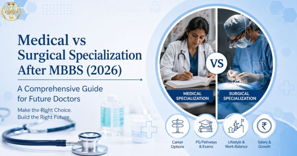
📅 Updated September 2026 ⏰ 9 min read 🎓 MBBS Graduates 🌎 Specialization Cluster — Blog 5 ⚡ Quick Answer Should I choose medical (MD) or surgical (MS) specialization after MBBS? Choosing between medical vs surgical specialization after MBBS comes down to temperament, not difficulty or prestige. If you enjoy diagnosing and managing conditions over time — often without an immediately visible endpoint — a physician-track (MD) branch usually fits better. If you’re drawn to procedures with a defined intervention and a faster feedback loop on outcomes, a surgical-track (MS) branch typically fits better. This is a different question from MD vs MS vs DNB, which compares qualification structure. This guide compares the clinical mindset each path actually demands. Deciding between medical vs surgical specialization after MBBS is more fundamental than picking a specific specialty: do you want to be the doctor who diagnoses and manages, or the doctor who operates? This decision — physician track versus surgical track — usually comes earlier than “which specific specialty,” and it shapes your entire training experience, day-to-day work pattern, and long-term career rhythm. This guide compares the two paths honestly: what physician training actually looks like day-to-day, what surgical training actually looks like, the stress profile each demands, and a set of reflective questions to help you figure out which one genuinely fits you — not which one sounds more impressive at a family gathering. Medical vs Surgical Specialization After MBBS: The Fundamental Question At its core, physician branches (MD) and surgical branches (MS) ask something different of you as a doctor. Physician medicine centres on diagnostic reasoning over time. You gather evidence, form a working diagnosis, start a treatment plan, and then watch how the patient responds — often adjusting your approach across multiple visits or admissions. The endpoint is frequently gradual: a chronic condition brought under control, a complex diagnosis eventually confirmed, a patient stabilised and monitored rather than “fixed” in a single moment. Surgery centres on decisive, procedural intervention. You assess, plan an operative approach, and then physically correct the problem — with a defined start and end to that specific intervention. The feedback loop is faster: you generally know within hours or days whether the operation achieved what it set out to do, even though full recovery and follow-up still take time. ℹ️ Neither mindset is “harder” or more valuable than the other. They’re different cognitive and emotional skill sets — sustained uncertainty tolerance versus decisive action under pressure — and most doctors are naturally more drawn to one than the other well before they can articulate why. This is the essence of the medical vs surgical specialization after MBBS decision. What Physician (MD) Training Actually Looks Like Day-to-Day A typical day in physician-track training centres on wards and outpatient clinics rather than an operating theatre. Morning ward rounds involve reviewing each patient’s overnight status, adjusting medications, and discussing evolving differential diagnoses with senior faculty. Outpatient department sessions bring a steady stream of new and follow-up patients, each requiring careful history-taking and clinical reasoning. Diagnostic work is central: interpreting lab results, imaging, and clinical signs together to build and refine a diagnosis, sometimes over several days or weeks as the picture becomes clearer. Treatment is rarely “one and done” — doctors titrate, monitor, and adjust it based on response. Physician trainees also typically carry a heavier academic and research load, since MD programmes place strong emphasis on dissertation work and structured case discussion. The physical demands are real but different from surgery — long hours on your feet during rounds, but less of the sustained standing and manual precision that operating requires. The mental demand is sustained: holding multiple evolving cases in your head simultaneously, often across different organ systems. What Surgical (MS) Training Actually Looks Like Day-to-Day Surgical training revolves around the operating theatre, though it involves far more than just operating. Pre-operative assessment — confirming diagnosis, planning the surgical approach, discussing risks with the patient — happens before every case. Trainees progress gradually from observing to assisting to performing increasing portions of procedures under supervision. Post-operative management is just as central as the operation itself: monitoring for complications, managing pain and wound care, and making judgment calls about when a patient is ready for discharge. Emergency and on-call surgical work adds genuine unpredictability — trauma, obstructed labour, or an acute abdomen doesn’t wait for a convenient time, and surgical training teaches residents to respond decisively regardless of the hour. Morbidity and mortality conferences are a defining feature of surgical training — structured, often uncomfortable discussions where teams review complications openly to improve future practice. Physically, surgical training is demanding in a specific way: extended periods standing during long operations, precise manual dexterity under time pressure, and the stamina to remain sharp through emergency cases at any hour. Medical vs Surgical Specialization After MBBS: Common MD and MS Branches Both tracks include a wide range of specific specialities, but a few branches illustrate the pattern clearly. MD — Physician Track Common MD Branches General Medicine, Dermatology, Radiology, Paediatrics, Psychiatry, Pathology, and Anaesthesiology. Focus: diagnosis, medical management, and longitudinal care within a defined system or across multiple systems. MS — Surgical Track Common MS Branches General Surgery, Orthopaedics, ENT (Otolaryngology), Obstetrics & Gynaecology, and Ophthalmology. Focus: operative correction, procedural skill, and defined clinical interventions. A few branches sit closer to the boundary — Obstetrics & Gynaecology, for instance, combines substantial medical management (antenatal care, hormonal conditions) with genuine operative work (deliveries, gynaecological surgery), which is worth considering if you’re drawn to elements of both tracks. Stress Profile & Decision-Making Style — Which Fits You? The medical vs surgical specialization after MBBS decision isn’t just about daily tasks. The two paths also demand different relationships with stress and decision-making. Physician-track stress tends to be sustained and diffuse. You’re often managing several evolving cases simultaneously, holding uncertainty for days or weeks while a diagnosis becomes clearer, and making incremental decisions that compound over time. The emotional weight comes from

Doctor examining a child representing common pediatric skin conditions in general practice
Learning blogs

Recognising Pediatric Skin Conditions: A Quick-Reference Guide for MBBS & Family Physicians

📅 Updated August 2026 ⏰ 8 min read 🩺 MBBS & Family Physicians 🌎 Dermatology Cluster — Day 6 ⚡ Quick Answer What are the most common pediatric skin conditions seen in Indian OPDs? Atopic dermatitis, miliaria (heat rash), diaper dermatitis, viral warts, insect-bite reactions, and nutritional-deficiency dermatoses are among the most frequently seen pediatric skin presentations in Indian general practice. Most respond well to basic measures. A smaller but important subset — rapidly spreading disease, signs of secondary infection, or atypical presentations — warrants pediatric dermatology referral. Pediatric skin conditions don’t behave like their adult counterparts. Treating them as though they do is one of the more common sources of misdiagnosis in general practice. Children present with a distinct set of conditions, at distinct ages, with distinct patterns. Parents bring a level of anxiety to a skin complaint that rarely accompanies an adult visiting with the same rash. This guide is a quick-reference for the pediatric skin conditions MBBS graduates and family physicians see most often in Indian OPDs. It covers what they look like and how they’re generally managed. Critically, it also covers when they warrant a referral rather than continued primary management. Why Pediatric Skin Conditions Need a Different Lens Children’s skin is structurally different from adult skin in ways that matter clinically. It’s thinner, has a higher surface-area-to-body-weight ratio, and its barrier function is still maturing — particularly in infants. The same irritant or infection can cause a mild, localised reaction in an adult. In a child, it can spread faster or present more diffusely. Age matters more in pediatric dermatology than in almost any other area of skin presentation. A rash that’s a normal, self-limiting finding in a two-week-old neonate can be an entirely different diagnosis in a five-year-old. Building a mental map of which conditions cluster at which ages — neonatal, infant, toddler, school-age — is genuinely useful. It’s one of the most useful diagnostic habits a general physician can develop. Finally, parental anxiety is a real clinical factor, not a distraction from the “real” medicine. A skin condition that’s medically minor can still generate significant parental distress. How a doctor communicates about it — timeline, expected course, what to watch for — shapes what happens next. It affects whether the family returns for appropriate follow-up or seeks multiple opinions out of worry. Atopic Dermatitis in Infants & Children Atopic dermatitis is the most common chronic skin condition seen in pediatric practice, and its presentation shifts meaningfully with age. Why atopic dermatitis occurs (pathophysiology) The pathophysiology of atopic dermatitis centres on two related problems. A defective skin barrier lets moisture escape and irritants in. An underlying immune system also overreacts to that irritation with inflammation, as a detailed review from the American Academy of Pediatrics lays out. This is part of why eczema occurs more readily in children with a family history of atopic conditions. That family history often includes asthma, allergic rhinitis, or atopic dermatitis itself. Understanding this pathogenesis helps explain why barrier repair, not just anti-inflammatory treatment, sits at the centre of long-term management. Age-pattern differences In infants, atopic dermatitis typically appears on the face, scalp, and extensor surfaces of the limbs. That’s a different distribution from the flexural pattern — elbows, knees — more familiar from adult and older-child presentations. As children grow, the distribution gradually shifts toward the flexural pattern seen in older children and adults. This age-based shift feeds into eczema differential diagnosis. It means distinguishing atopic dermatitis from miliaria, contact dermatitis, or an early fungal infection. Our companion guide on eczema, psoriasis, and fungal rash differentiation covers this in more depth. General management principles Consistent moisturising with a fragrance-free emollient is the foundation of management at any age. Barrier repair reduces flare frequency more reliably than treating flares alone. Identifying and minimising specific triggers — harsh soaps, wool fabric, prolonged hot baths — also helps. Many families reduce flare frequency this way without needing additional intervention. This combination — consistent barrier care plus trigger avoidance — is the most effective eczema prevention approach at the primary-care level. It works better than treating flares reactively as they appear. When to refer Refer when atopic dermatitis is extensive, unresponsive to basic measures, or shows signs of infected eczema. Oozing, honey-coloured crusting, or fever accompanying a flare are the classic markers of secondary bacterial infection. Persistent sleep disruption from itching in a young child is also a reasonable trigger for earlier specialist involvement. That’s given the impact on the child’s wellbeing and family quality of life. Miliaria & Diaper Dermatitis: Managing the Basics Miliaria (heat rash) Extremely common in Indian infants given the climate, miliaria results from sweat duct obstruction. It presents as small clear or red bumps, typically on the neck, chest, and skin folds. Cooling measures — lighter clothing, reduced overheating, staying in ventilated spaces during peak heat — are usually enough. Active treatment usually isn’t needed. It usually resolves once the underlying heat and sweating trigger is addressed. Diaper dermatitis Most diaper dermatitis is irritant contact dermatitis from prolonged exposure to moisture and friction. It responds to basic hygiene measures — frequent diaper changes, gentle cleansing, and adequate drying time between changes. The distinction worth knowing is candidal diaper dermatitis, which presents differently. It shows a bright red rash with well-demarcated edges and satellite lesions. Those lesions are small separate red spots surrounding the main rash, extending into the skin folds. This pattern suggests a fungal component rather than simple irritation. It generally needs a different management approach than basic hygiene alone. ℹ️ Diaper dermatitis that doesn’t improve with consistent basic hygiene measures after a reasonable trial is worth reconsidering. The same applies if it shows the satellite-lesion pattern — that’s a cue to rethink the diagnosis. Viral Warts, Insect Bites & Common Infections Viral warts Common in school-age children, HPV-caused viral warts present as rough, well-defined papules, often on the hands and feet. They’re generally self-limited over time but can persist for months to years. They

Doctors in a modern hospital representing the future scope of medical specialization in India
Learning blogs

Future Scope of Medical Specialization in India: What the Next Decade Looks Like

📅 Updated August 2026 ⏰ 9 min read 🎓 MBBS Students & Junior Doctors 🌎 Specialization Cluster — Trend Guide ⚡ Quick Answer Which medical specializations have the strongest future scope in India? Critical Care, Radiology, Fetal Medicine, Oncology, Diabetology, and Emergency Medicine are structurally positioned for sustained demand over the next decade, driven by India’s ageing population, its rising chronic disease burden, and the expansion of diagnostic and tertiary care infrastructure. Structural demand is a useful input into your specialty decision — but it should complement genuine clinical interest, not replace it. If you’re choosing a specialty today, you’re not just choosing what to practice next year. You’re choosing what you’ll be doing in 2040 and beyond. That’s a long horizon, and it’s reasonable to ask which fields are likely to still have strong demand decades from now. That includes adequate infrastructure and career relevance too — not just which specialty is competitive on today’s NEET PG cutoffs. This guide looks at the future scope of medical specialization in India. It examines the structural forces reshaping India’s healthcare needs — demographic change, disease burden shift, and technology adoption. It also includes an honest caution: future scope is one input into a good decision, not the whole decision. What Drives the Future Scope of Medical Specialization in India Current-day demand and structural, multi-decade demand are not the same thing. A specialty can be competitive today because of a temporary hiring wave, a popular perception, or a limited number of seats. None of that says much about whether the demand will hold for the next 20 years. Structural demand, by contrast, is driven by forces that move slowly but predictably. Those forces are who the population will be, what diseases they’ll carry, and what infrastructure will exist to treat them. Three forces matter most for India specifically: Demographic shift — how the age structure of the population is changing, and what that means for disease patterns Disease burden shift — the ongoing move from a primarily infectious-disease burden toward a growing non-communicable and chronic disease burden Infrastructure and technology expansion — the growth of tertiary care, diagnostic capacity, and digital health systems. This makes certain kinds of care more accessible and more in demand None of these forces guarantees an individual doctor’s success in a given field. That still depends on skill, institution, and location. But they do indicate where the underlying patient need is heading. That’s a meaningfully different question from where the current hiring market happens to be. Demographic Shift: India’s Ageing Population & Chronic Disease Burden India’s ageing population India is often described by its youthful demographic — and that’s accurate today. But the same population is ageing at a pace that will reshape healthcare demand. That reshaping will happen within the working lifetime of anyone graduating MBBS now. According to the UNFPA India Ageing Report 2023, India’s population aged 60 and above is projected to grow substantially by 2050. It’s expected to rise from 149 million in 2022 to 347 million by 2050. The share of elderly is also expected to rise, from around 10.5% to over 20% of the total population. By 2046, the report projects that the elderly population will actually surpass the population of children aged 0–15. The 80-and-above age group specifically is projected to grow by around 279% over the same period. That group typically carries the highest burden of multi-morbidity and dependency. This shift has direct clinical implications. An ageing population means more patients with multiple concurrent conditions. It also means more need for critical care and geriatric-oriented management, and rising demand for oncology, cardiology, and rehabilitation services. Rising chronic disease burden Alongside ageing, India’s chronic disease burden is already substantial and growing. The ICMR-INDIAB national study, published in The Lancet Diabetes & Endocrinology in 2023, estimated 101 million people living with diabetes across India. It also found a further 136 million with prediabetes and an estimated 315 million with hypertension. These are not projections of a future problem — this is the current disease burden. It’s expected to keep rising as risk factors like obesity and sedentary lifestyles increase. ℹ️ An ageing population and a large, growing diabetic and hypertensive population together create compounding demand. That demand spans several specialties at once — not just endocrinology, but cardiology, nephrology, and ophthalmology for diabetic retinopathy. It also spans critical care, since uncontrolled chronic disease is a major driver of ICU admissions. Technology Shift: Imaging, Diagnostics & Digital Health The second structural driver is less about who patients are and more about how care is delivered. India’s diagnostic and digital health infrastructure has been expanding steadily, and this expansion changes which specialties see rising demand. Diagnostic infrastructure growth As tertiary care hospitals and diagnostic chains expand into smaller cities, imaging, laboratory, and referral volumes all grow. This directly benefits imaging- and lab-based specialties. Every new CT or MRI installation, every new diagnostic centre, represents ongoing demand for radiologists and pathologists to interpret the data. Digital health and teleconsultation Government digital health initiatives and the broader adoption of telemedicine have made remote specialist consultation far more feasible. Reporting is also far easier than it was a decade ago. This particularly benefits specialties where the core clinical work is interpretive rather than procedural at the bedside. Radiology and pathology again stand out, since scan and slide interpretation can be done remotely. That expands the geographic reach of a single specialist. AI-assisted diagnostics AI-assisted tools are used to support — not replace — radiologists and pathologists, helping triage and flag findings for review. Rather than reducing demand for these specialists, the practical effect so far has been different. It has helped manage a rapidly growing volume of studies that outpaces the number of specialists available to read them. Prenatal screening and genetic testing Growing awareness and access to prenatal screening and genetic testing has expanded demand for fetal medicine. That specialty combines advanced imaging with genetic counselling for high-risk pregnancies. It remains under-supplied relative to

Doctors discussing hospital roles representing RMO vs duty doctor vs junior resident careers
Learning blogs

RMO vs Duty Doctor vs Junior Resident: Which Builds a Better Career After MBBS?

Every year, roughly a lakh MBBS graduates in India finish their internship and face the same first decision: which job to take next. Three titles come up constantly in this search — Resident Medical Officer (RMO), Duty Doctor, and Junior Resident (JR) — and they’re routinely used as if they mean the same thing. They don’t, and the difference matters more than most graduates realise when they’re signing their first offer letter. This guide breaks down what each role actually involves in Indian private hospitals and nursing homes, how they compare on pay, hours, and clinical learning, and — more importantly — what actually determines whether a role builds your career, because it usually isn’t the job title on the offer letter. Quick Answer If your priority is NEET-PG or a future specialisation, a Junior Resident post in a teaching hospital generally offers the strongest structured foundation — supervised procedures, senior-led case discussions, and academic integration. If your priority is immediate income, general clinical exposure, or time to decide your next step, an RMO or Duty Doctor role in a private hospital is a reasonable and often more accessible choice. But the title alone won’t tell you which one you’re getting. The same title means very different things at different hospitals — what actually determines the value of the role is covered below. Understanding the Three Roles All three roles are held by qualified, registered medical graduates who provide direct patient care in a hospital setting. Where they diverge is in training status, institutional setting, and how the role is structured around (or outside of) formal postgraduate education. Resident Medical Officer (RMO) An RMO is a full-time, salaried doctor employed to provide continuous, round-the-clock inpatient coverage — most commonly in private hospitals, corporate chains, and nursing homes. The role is service-oriented rather than academic: it exists to keep a doctor physically present in the hospital at all times, acting as the link between nursing staff, admitted patients, and visiting or on-call consultants. Duty Doctor This is where most of the confusion starts. “Duty Doctor” isn’t really a separate employment category — it’s a functional title for whichever doctor is currently assigned to cover the OPD, a ward, or the casualty unit during a specific shift block. In practice, an RMO or a Junior Resident can be the “duty doctor” for their assigned shift. Some smaller hospitals and nursing homes use “Duty Doctor” as their standard job title for what is functionally an RMO position; others use it strictly to mean whoever is on casualty/emergency coverage right now, regardless of their formal designation. Junior Resident (JR) A Junior Resident has completed MBBS and internship and is working within a structured departmental training post — most often in a teaching hospital, medical college, or large private hospital running an academic programme. JRs fall into two categories: academic JRs, formally enrolled in a postgraduate degree (MD/MS/DNB) while training, and non-academic JRs, working in a similar structured clinical role without current postgraduate enrolment. Either way, the defining feature is supervision within a department, not just a shift roster. Where these sit in the hierarchy: Intern → Junior Resident → Senior Resident → Consultant is the academic training ladder most teaching hospitals follow. RMO and Duty Doctor roles generally sit outside this ladder, as a parallel service-employment track most common in non-teaching private hospitals and nursing homes. Role-by-Role Breakdown: Responsibilities & Work Pattern Here’s what each role typically looks like day-to-day. Aspect RMO Duty Doctor Junior Resident Typical setting Private hospitals, nursing homes, corporate chains Same as RMO — a shift assignment, not a separate setting Teaching hospitals, medical colleges, academic private hospitals Core focus Continuous ward coverage, patient monitoring, admin Immediate response for whoever is assigned that shift/unit Clinical service combined with structured academic training Typical duties Ward rounds, vitals monitoring, admissions, initial emergency response, documentation OPD consults, ward rounds, casualty triage — depending on the assigned unit Ward admissions, assisting in procedures/surgery, bedside procedures, case presentations Supervision Reports to hospital management and on-call/visiting consultants Same as whichever role (RMO/JR) is holding the assignment Senior Residents and unit consultants/professors Shift pattern Fixed rotating shifts — commonly 8 or 12 hours Defined by the shift block being covered Longer, less predictable hours, including frequent 24-hour on-call stretches Notice that “Duty Doctor” doesn’t get its own distinct row for supervision or setting — that’s intentional. It genuinely is a shift-based label layered on top of an RMO or JR position at most hospitals, not a fourth category with its own separate structure. Pay & Work-Hours Comparison Fresher-level compensation across India commonly falls in the ₹40,000–₹90,000/month range, with meaningful variation by city, hospital tier, and department. Where the three roles differ more consistently is not the absolute number but the structure of the pay itself. RMO: Usually a fixed monthly salary tied to a defined shift roster. Predictable income, predictable hours on paper — though rotating night shifts still take a toll. Duty Doctor: Sometimes fixed-salary (when it’s simply another name for the RMO post at that hospital), sometimes paid per shift or on a locum basis — particularly common in smaller nursing homes that need flexible casualty coverage without a full-time hire. Junior Resident: Typically a fixed monthly stipend set by the hospital or university’s residency pay scale. Because the role combines training with service, the effective hourly rate is often lower than it looks on paper, given the frequency of extended on-call hours. Before accepting any offer, ask directly whether the pay is fixed or shift-based, what the expected weekly hours actually are (not just the “official” shift length), and whether night duty or on-call time is compensated separately. Compensation figures referenced here are based on general fresher-level ranges observed across India and are not specific to any single role, hospital, or city. Actual pay varies significantly by location, hospital tier, and individual negotiation — always confirm exact figures directly with the hospital before accepting an offer. Which Offers the Most Clinical Learning? This

Scroll to Top